Luders Eileen, Di Paola Margherita, Tomaiuolo Francesco, Thompson Paul M, Toga Arthur W, Vicari Stefano, Petrides Michael, Caltagirone Carlo
Laboratory of Neuro Imaging, Department of Neurology, UCLA School of Medicine, Los Angeles, California 90095-7334, USA, and IRCCS Ospedale Pediatrico Bambino Gesu, Roma, Italy.
Neuroreport. 2007 Feb 12;18(3):203-7. doi: 10.1097/WNR.0b013e3280115942.
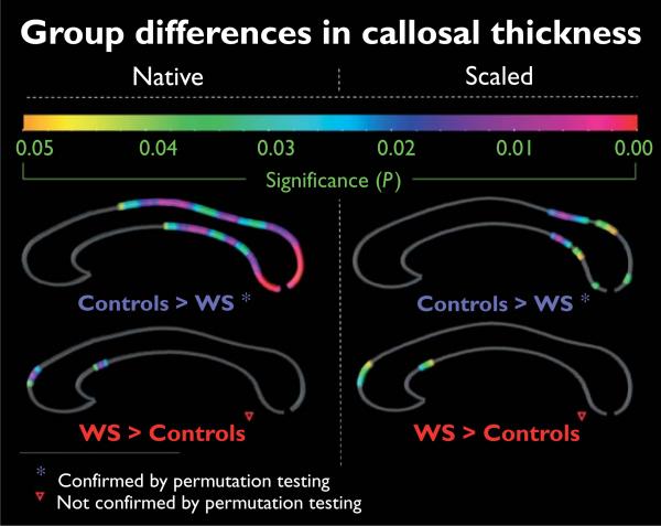
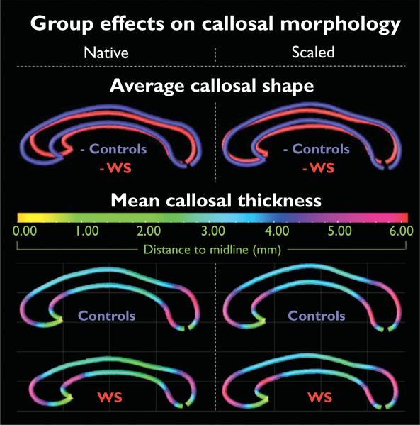
We applied novel mesh-based geometrical modeling methods to calculate and compare the thickness of the corpus callosum at high spatial resolution and to create profiles of average callosal shape in a well-matched sample (n=24) of individuals with Williams syndrome and controls. In close agreement with previous observations, superimposed surface maps indicate that the corpus callosum in Williams syndrome individuals is shorter and less curved. Moreover, we observed significantly thinner callosal regions in Williams syndrome individuals across the posterior surface, where group effects were less pronounced and spatially restricted in brain-size-adjusted data compared with native data. Circumscribed structural alterations in callosal morphology might be candidate anatomic substrates for the unique cognitive and behavioral profile associated with Williams syndrome.
我们应用了基于新型网格的几何建模方法,以高空间分辨率计算并比较胼胝体的厚度,并在威廉姆斯综合征患者和对照组的匹配良好的样本(n = 24)中创建平均胼胝体形状的剖面图。与先前的观察结果密切一致,叠加的表面图表明威廉姆斯综合征患者的胼胝体更短且弯曲度更小。此外,我们观察到威廉姆斯综合征患者后表面的胼胝体区域明显更薄,与原始数据相比,在脑大小调整后的数据中,组间效应不那么明显且在空间上受到限制。胼胝体形态的局限性结构改变可能是与威廉姆斯综合征相关的独特认知和行为特征的候选解剖学基础。