• 文献检索
  • 文档翻译
  • 深度研究
  • 学术资讯
  • Suppr Zotero 插件Zotero 插件
  • 邀请有礼
  • 套餐&价格
  • 历史记录
应用&插件
Suppr Zotero 插件Zotero 插件浏览器插件Mac 客户端Windows 客户端微信小程序
定价
高级版会员购买积分包购买API积分包
服务
文献检索文档翻译深度研究API 文档MCP 服务
关于我们
关于 Suppr公司介绍联系我们用户协议隐私条款
关注我们

Suppr 超能文献

核心技术专利:CN118964589B侵权必究
粤ICP备2023148730 号-1Suppr @ 2026

文献检索

告别复杂PubMed语法,用中文像聊天一样搜索,搜遍4000万医学文献。AI智能推荐,让科研检索更轻松。

立即免费搜索

文件翻译

保留排版,准确专业,支持PDF/Word/PPT等文件格式,支持 12+语言互译。

免费翻译文档

深度研究

AI帮你快速写综述,25分钟生成高质量综述,智能提取关键信息,辅助科研写作。

立即免费体验

霍奇金里德-斯腾伯格细胞中c-fos/c-jun表达与组织细胞分化的相关性。对具有自发分化能力的HDLM-1亚克隆进行检测。

Correlation of c-fos/c-jun expression with histiocytic differentiation in Hodgkin's Reed-Sternberg cells. Examination in HDLM-1 subclones with spontaneous differentiation.

作者信息

Hsu S M, Xie S S, el-Okda M O, Hsu P L

机构信息

Department of Pathology, University of Arkansas for Medical Sciences, Little Rock.

出版信息

Am J Pathol. 1992 Jan;140(1):155-65.

PMID:1731522
原文链接:https://pmc.ncbi.nlm.nih.gov/articles/PMC1886249/
Abstract

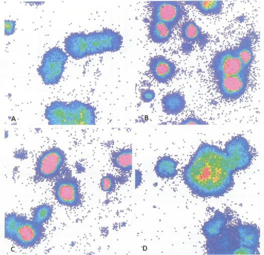
The c-fos proto-oncogene, which is the normal homolog of the transforming gene carried by murine osteogenic sarcoma viruses, interacts with the protein product of another proto-oncogene, c-jun, to form a heterodimer that can recognize and bind to a specific sequence of nucleotides in the DNA. The expression of c-fos and c-jun is linked to the proliferation of certain cells and the differentiation of others, including those of monomyelocyte lineage. The authors used two cultured Hodgkin's Reed-Sternberg (H-RS) cell lines, KM-H2 and HDLM-1, and their single-cell clones to study the correlation of c-fos/c-jun expression with cell differentiation in H-RS cells. Within 48 hours after induction with phorbol ester (TPA), both parent lines exhibited markedly increased expression of c-fos/c-jun. The expression returned to the preinduction level after 96 hours, however, and the cells retained their differentiated status. The transitory increase in c-fos/c-jun expression suggests that binding of these proteins to a specific promoter in the nucleus triggers a cascade of events that result in cell differentiation. Expression of these proteins may not be required for the cells to maintain their differentiation. The authors selected three groups of sublines of HDLM-1 cells based on their degree of spontaneous cytologic differentiation. The first group, without obvious differentiation, showed a c-fos/c-jun expression pattern similar to that of the parent line. The second group, with moderate differentiation, had a high degree of expression, which decreased on treatment with TPA. The third group, which had morphologic features resembling those of histiocytes, expressed minimal amounts of c-fos/c-jun, irrespective of TPA treatment. These findings provide further evidence that c-fos/c-jun expression is related to differentiation of H-RS cells, and that these proteins are not byproducts of TPA induction. Expression of c-fos/c-jun also was noted in a subpopulation of H-RS cells in tissues; and this expression also was enhanced when these cells were treated with TPA in culture. These findings indicate that H-RS cells can differentiate to become mature-appearing cells in tissues.

摘要

c-fos原癌基因是鼠成骨肉瘤病毒携带的转化基因的正常同源物,它与另一个原癌基因c-jun的蛋白质产物相互作用,形成一个异二聚体,该异二聚体能够识别并结合DNA中的特定核苷酸序列。c-fos和c-jun的表达与某些细胞的增殖以及其他细胞(包括单核细胞系细胞)的分化有关。作者使用两种培养的霍奇金里德-斯腾伯格(H-RS)细胞系KM-H2和HDLM-1及其单细胞克隆,来研究H-RS细胞中c-fos/c-jun表达与细胞分化的相关性。在用佛波酯(TPA)诱导后的48小时内,两个亲代细胞系均表现出c-fos/c-jun表达显著增加。然而,96小时后表达恢复到诱导前水平,且细胞保持其分化状态。c-fos/c-jun表达的短暂增加表明,这些蛋白质与细胞核中特定启动子的结合触发了一系列导致细胞分化的事件。这些蛋白质的表达可能不是细胞维持其分化所必需的。作者根据HDLM-1细胞的自发细胞学分化程度选择了三组亚系。第一组没有明显分化,其c-fos/c-jun表达模式与亲代细胞系相似。第二组有中度分化,表达程度高,用TPA处理后表达降低。第三组具有类似于组织细胞的形态特征,无论是否用TPA处理,均表达极少量的c-fos/c-jun。这些发现进一步证明c-fos/c-jun表达与H-RS细胞的分化有关,且这些蛋白质不是TPA诱导的副产物。在组织中的H-RS细胞亚群中也观察到了c-fos/c-jun的表达;当这些细胞在培养中用TPA处理时,这种表达也会增强。这些发现表明,H-RS细胞在组织中可以分化为外观成熟的细胞。

https://cdn.ncbi.nlm.nih.gov/pmc/blobs/5469/1886249/6039409bd51a/amjpathol00085-0163-a.jpg
https://cdn.ncbi.nlm.nih.gov/pmc/blobs/5469/1886249/8cca0a524997/amjpathol00085-0159-a.jpg
https://cdn.ncbi.nlm.nih.gov/pmc/blobs/5469/1886249/02af02dd914c/amjpathol00085-0160-a.jpg
https://cdn.ncbi.nlm.nih.gov/pmc/blobs/5469/1886249/6f26a5134bd6/amjpathol00085-0161-a.jpg
https://cdn.ncbi.nlm.nih.gov/pmc/blobs/5469/1886249/6039409bd51a/amjpathol00085-0163-a.jpg

相似文献

1
Correlation of c-fos/c-jun expression with histiocytic differentiation in Hodgkin's Reed-Sternberg cells. Examination in HDLM-1 subclones with spontaneous differentiation.霍奇金里德-斯腾伯格细胞中c-fos/c-jun表达与组织细胞分化的相关性。对具有自发分化能力的HDLM-1亚克隆进行检测。
Am J Pathol. 1992 Jan;140(1):155-65.
2
Cultured Reed-Sternberg cells HDLM-1 and KM-H2 can be induced to become histiocytelike cells. H-RS cells are not derived from lymphocytes.培养的里德-斯腾伯格细胞HDLM-1和KM-H2可被诱导成为组织细胞样细胞。霍奇金里德-斯腾伯格细胞并非来源于淋巴细胞。
Am J Pathol. 1990 Aug;137(2):353-67.
3
Reed-Sternberg cells in Hodgkin's cell lines HDLM, L-428, and KM-H2 are not actively replicating: lack of bromodeoxyuridine uptake by multinuclear cells in culture.霍奇金细胞系HDLM、L-428和KM-H2中的里德-斯腾伯格细胞并非在活跃复制:培养中的多核细胞缺乏溴脱氧尿苷摄取。
Blood. 1988 May;71(5):1382-9.
4
Expression of macrophage colony-stimulating factor (M-CSF) in two Hodgkin's Reed-Sternberg (H-RS) cell lines, HDLM-1 and KM-H2, and in H-RS cells in tissues.巨噬细胞集落刺激因子(M-CSF)在两种霍奇金里德-斯腾伯格(H-RS)细胞系HDLM-1和KM-H2以及组织中的H-RS细胞中的表达。
Int J Hematol. 1991 Aug;54(4):315-26.
5
Lack of effect of colony-stimulating factors, interleukins, interferons, and tumor necrosis factor on the growth and differentiation of cultured Reed-Sternberg cells. Comparison with effects of phorbol ester and retinoic acid.集落刺激因子、白细胞介素、干扰素及肿瘤坏死因子对培养的里德-斯腾伯格细胞生长和分化无作用。与佛波酯和视黄酸作用的比较。
Am J Pathol. 1990 Jan;136(1):181-9.
6
Expression of cytokine genes, cytokine receptor genes, and transcription factors in cultured Hodgkin and Reed-Sternberg cells.培养的霍奇金和里德-斯腾伯格细胞中细胞因子基因、细胞因子受体基因及转录因子的表达
Cancer Res. 1992 Jun 15;52(12):3353-60.
7
Phenotypes and phorbol ester-induced differentiation of human histiocytic lymphoma cell lines (U-937 and SU-DHL-1) and Reed-Sternberg cells.人组织细胞淋巴瘤细胞系(U-937和SU-DHL-1)及里德-施特恩伯格细胞的表型和佛波酯诱导的分化
Am J Pathol. 1986 Feb;122(2):223-30.
8
Fos and Jun form cell specific protein complexes at the neuropeptide tyrosine promoter.Fos和Jun在神经肽酪氨酸启动子处形成细胞特异性蛋白复合物。
Oncogene. 1994 Aug;9(8):2369-75.
9
Differential induction of c-fos and c-jun proto-oncogenes and AP-1 activity by tumor promoter 12-O-tetradecanoyl phorbol 13-acetate in cells at different stages of tumor promotion in vitro.肿瘤促进剂12-O-十四烷酰佛波醇-13-乙酸酯在体外肿瘤促进不同阶段的细胞中对c-fos和c-jun原癌基因的差异诱导及AP-1活性
Oncogene. 1992 Nov;7(11):2287-94.
10
Identification of an Mr 70,000 antigen associated with Reed-Sternberg cells and interdigitating reticulum cells.与里德-施特恩伯格细胞和交错突网状细胞相关的一种分子量为70,000的抗原的鉴定。
Cancer Res. 1990 Jan 15;50(2):350-7.

引用本文的文献

1
Hodgkin lymphoma cell lines bind to platelets. Incubation with platelets induces CD15 and P-selectin dependent adhesion of the cell lines to Human Umbilical Vein Endothelial cells (HUVEC).霍奇金淋巴瘤细胞系与血小板结合。与血小板共同孵育可诱导细胞系通过CD15和P选择素依赖的方式黏附于人脐静脉内皮细胞(HUVEC)。
Cancer Biol Ther. 2015;16(11):1651-9. doi: 10.1080/15384047.2015.1095411. Epub 2015 Sep 29.
2
AP-1 mediated relief of repressive activity of the CD30 promoter microsatellite in Hodgkin and Reed-Sternberg cells.AP-1介导霍奇金和里德-斯腾伯格细胞中CD30启动子微卫星抑制活性的解除。
Am J Pathol. 2003 Aug;163(2):633-41. doi: 10.1016/S0002-9440(10)63690-5.
3

本文引用的文献

1
Production of a monoclonal antibody specific for Hodgkin and Sternberg-Reed cells of Hodgkin's disease and a subset of normal lymphoid cells.制备一种针对霍奇金病的霍奇金细胞和施特恩伯格-里德细胞以及一部分正常淋巴细胞的单克隆抗体。
Nature. 1982 Sep 2;299(5878):65-7. doi: 10.1038/299065a0.
2
Color modification of diaminobenzidine (DAB) precipitation by metallic ions and its application for double immunohistochemistry.金属离子对二氨基联苯胺(DAB)沉淀的颜色修饰及其在双重免疫组织化学中的应用。
J Histochem Cytochem. 1982 Oct;30(10):1079-82. doi: 10.1177/30.10.6182185.
3
Use of avidin-biotin-peroxidase complex (ABC) in immunoperoxidase techniques: a comparison between ABC and unlabeled antibody (PAP) procedures.
Regulation of fos-lacZ fusion gene expression in primary mouse epidermal keratinocytes isolated from transgenic mice.
从转基因小鼠分离的原代小鼠表皮角质形成细胞中fos-lacZ融合基因表达的调控
Biochem J. 1994 May 15;300 ( Pt 1)(Pt 1):263-70. doi: 10.1042/bj3000263.
抗生物素蛋白-生物素-过氧化物酶复合物(ABC)在免疫过氧化物酶技术中的应用:ABC法与未标记抗体(PAP)法的比较。
J Histochem Cytochem. 1981 Apr;29(4):577-80. doi: 10.1177/29.4.6166661.
4
Phenotypic expression of Hodgkin's and Reed-Sternberg cells in Hodgkin's disease.霍奇金病中霍奇金和里德-斯腾伯格细胞的表型表达。
Am J Pathol. 1985 Feb;118(2):209-17.
5
Rapid induction of the expression of proto-oncogene fos during human monocytic differentiation.人单核细胞分化过程中原癌基因fos表达的快速诱导。
Cell. 1985 Jan;40(1):209-17. doi: 10.1016/0092-8674(85)90324-1.
6
Hodgkin's disease derived cell lines HDLM-2 and L-428: comparison of morphology, immunological and isoenzyme profiles.
Leuk Res. 1986;10(5):487-500. doi: 10.1016/0145-2126(86)90084-6.
7
Extracellular matrix does not induce the proliferation, but promotes the differentiation, of Hodgkin's cell line HDLM-1.细胞外基质不会诱导霍奇金细胞系HDLM-1的增殖,但会促进其分化。
Am J Pathol. 1987 Apr;127(1):9-14.
8
Expression of interleukin-1 in Reed-Sternberg cells and neoplastic cells from true histiocytic malignancies.白细胞介素-1在里德-斯腾伯格细胞及真性组织细胞性恶性肿瘤的肿瘤细胞中的表达。
Am J Pathol. 1986 Nov;125(2):221-5.
9
Phenotypes and phorbol ester-induced differentiation of human histiocytic lymphoma cell lines (U-937 and SU-DHL-1) and Reed-Sternberg cells.人组织细胞淋巴瘤细胞系(U-937和SU-DHL-1)及里德-施特恩伯格细胞的表型和佛波酯诱导的分化
Am J Pathol. 1986 Feb;122(2):223-30.
10
c-fos oncogene expression in human hematopoietic malignancies is restricted to acute leukemias with monocytic phenotype and to subsets of B cell leukemias.原癌基因c-fos在人类造血系统恶性肿瘤中的表达仅限于具有单核细胞表型的急性白血病以及B细胞白血病的某些亚群。
Blood. 1987 Nov;70(5):1450-7.