Mainero Caterina, Zhang Wei-Ting, Kumar Ashok, Rosen Bruce R, Sorensen A Gregory
Athinoula A. Martinos Center for Biomedical Imaging, Massachusetts General Hospital, Harvard Medical School, Charlestown, MA 02129, USA.
Neuroimage. 2007 Apr 15;35(3):1201-10. doi: 10.1016/j.neuroimage.2007.01.024. Epub 2007 Feb 4.
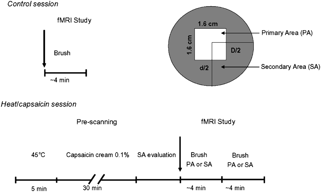
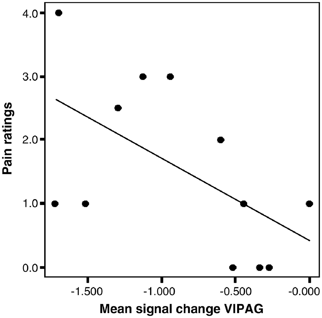
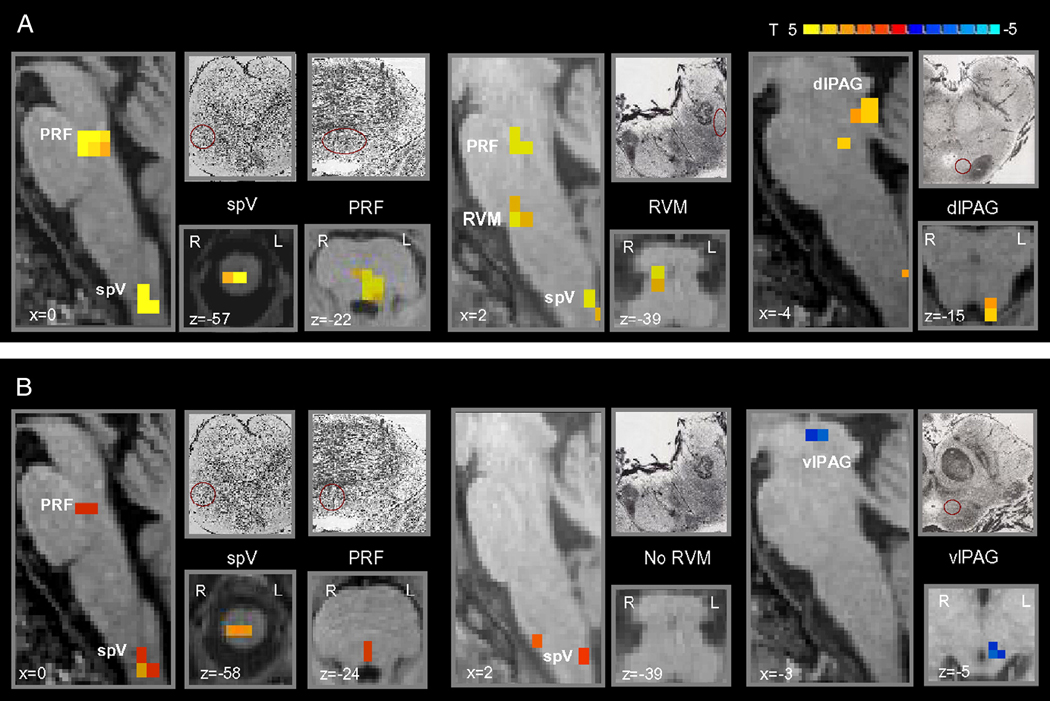
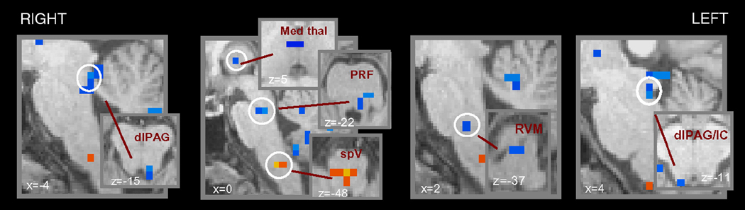
Following injury and inflammation, pain to light stroking (dynamic mechanical allodynia) might develop at the damaged site (primary area) or in adjacent normal tissue (secondary area). Using fMRI we mapped changes in the spinal trigeminal nucleus (spV), and supraspinal brainstem nuclei following heat/capsaicin-induced primary and secondary dynamic mechanical allodynia in the human trigeminal system. The role of these structures in dynamic mechanical allodynia has not been clarified yet in humans. During the control session we applied the same mechanical stimuli to the same untreated trigeminal area. Primary and secondary mechanical allodynia showed equal levels of perceived pain intensity, and compared to control mechanical stimulation exhibited similar responses in the ipsilateral spV and contralateral ventrolateral periaqueductal gray (vlPAG). Activity in the spV was significantly higher during both conditions versus the control mechanical stimulation, indicating that central sensitization of second-order neurons is similar for primary and secondary mechanical allodynia. The vlPAG showed decreased activity that inversely correlated with pain ratings during primary allodynia, i.e. the more deactivated the vlPAG the higher the pain intensity (p<0.05, Pearson's correlation). Primary and secondary dynamic mechanical allodynia were also characterized by significant differences involving distinct supraspinal structures mainly involved in pain modulation and including the rostroventromedial medulla, pons reticular formation, dorsolateral PAG, all more active during primary versus secondary allodynia, and the medial reticular formation of the caudal medulla that was more active during secondary versus primary allodynia. These results indicate that the pain modulatory system is involved to a different extent during primary versus secondary mechanical allodynia.
在损伤和炎症后,受损部位(原发区域)或相邻正常组织(继发区域)可能会出现对轻触的疼痛(动态机械性异常性疼痛)。我们使用功能磁共振成像(fMRI)绘制了热/辣椒素诱导的人类三叉神经系统原发性和继发性动态机械性异常性疼痛后,三叉神经脊束核(spV)和脊髓上脑干核的变化。这些结构在动态机械性异常性疼痛中的作用在人类中尚未明确。在对照阶段,我们对相同未处理的三叉神经区域施加相同的机械刺激。原发性和继发性机械性异常性疼痛表现出相同水平的疼痛强度感知,并且与对照机械刺激相比,在同侧spV和对侧腹外侧导水管周围灰质(vlPAG)中表现出相似的反应。与对照机械刺激相比,在两种情况下spV中的活动均显著更高,表明原发性和继发性机械性异常性疼痛中二级神经元的中枢敏化相似。vlPAG在原发性异常性疼痛期间表现出与疼痛评分呈负相关的活动降低,即vlPAG失活越明显,疼痛强度越高(p<0.05,Pearson相关性)。原发性和继发性动态机械性异常性疼痛的特征还在于涉及主要参与疼痛调节的不同脊髓上结构的显著差异,包括吻侧腹内侧延髓、脑桥网状结构、背外侧导水管周围灰质,在原发性与继发性异常性疼痛期间均更活跃,以及尾侧延髓的内侧网状结构在继发性与原发性异常性疼痛期间更活跃。这些结果表明,在原发性与继发性机械性异常性疼痛期间,疼痛调节系统的参与程度不同。