Kansai College of Oriental Medicine, 2-11-1 Wakaba Kumatori-cho Sennan-gun, Osaka 590-0482, Department of Pharmacology, Medicine Kinki University School of Medicine 377-2 Ohno-Higashi, Osaka-Sayama, Osaka 589-8511 and Department of Science, Pip-Fujimoto Co. Ltd, 1-36 Noninbashi 2-choume, Chuo-ku, Osaka 540-0011, Japan.
Evid Based Complement Alternat Med. 2007 Mar;4(1):99-105. doi: 10.1093/ecam/nel067. Epub 2006 Nov 2.
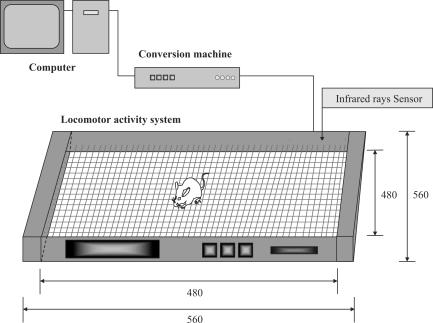
In order to examine the effectiveness of applying a static magnetic field (SMF) for increasing bone mineral density (BMD), we assessed the degree of osteopenia by dual-energy X-ray absorptiometry (DEXA), the metabolism measuring system, and histological examination of bone tissue in an ovariectomized (OVX) rat model. Thirty-six female Wistar rats (8 weeks old, 160-180 g) were divided into three groups. The rats in the OVX-M group were exposed to SMF for 12 weeks after ovariectomy. The ovariectomized rats in the OVX-D group were not exposed to SMF as a control. The rats in the normal group received neither ovariectomy nor exposure to SMF. Twelve-week exposure to SMF in the OVX-M group inhibited the reduction in BMD that was observed in the OVX-D group. Moreover, in the OVX rats, before exposure to SMF, there was no clear difference in the level of locomotor activity between the active and resting phases, and the pattern of locomotor activity was irregular. After exposure of OVX rats to SMF, the pattern of locomotor activity became diphasic with clear active and resting phases, as was observed in the normal group. In the OVX-M group, the continuity of the trabecular bone was maintained more favorably and bone mass was higher than the respective parameters in the OVX-D group. These results demonstrate that exposure to SMF increased the level of locomotor activity in OVX rats, thereby increasing BMD.
为了研究静磁场(SMF)应用于增加骨矿物质密度(BMD)的效果,我们使用双能 X 射线吸收法(DEXA)、代谢测量系统和骨组织的组织学检查评估了去卵巢(OVX)大鼠模型中的骨质减少程度。将 36 只雌性 Wistar 大鼠(8 周龄,160-180g)分为三组。OVX-M 组大鼠在去卵巢后接受 12 周的 SMF 照射。OVX-D 组的去卵巢大鼠未接受 SMF 照射作为对照。正常组大鼠既未接受去卵巢手术也未接受 SMF 照射。在 OVX-M 组中,12 周的 SMF 照射抑制了 OVX-D 组中观察到的 BMD 降低。此外,在接受 SMF 照射之前,未去卵巢的大鼠在活动和休息阶段的运动活性水平没有明显差异,运动活性模式不规则。在 OVX 大鼠暴露于 SMF 后,运动活性模式变得双相,具有明显的活动和休息阶段,与正常组相似。在 OVX-M 组中,小梁骨的连续性更有利地保持,骨量高于 OVX-D 组的相应参数。这些结果表明,SMF 暴露增加了 OVX 大鼠的运动活性水平,从而增加了 BMD。