Gowen E, Miall R C
Faculty of Life Sciences, Moffat Building, The University of Manchester, Manchester, M60 1QD, UK.
Neuroimage. 2007 Jun;36(2):396-410. doi: 10.1016/j.neuroimage.2007.03.005. Epub 2007 Mar 20.
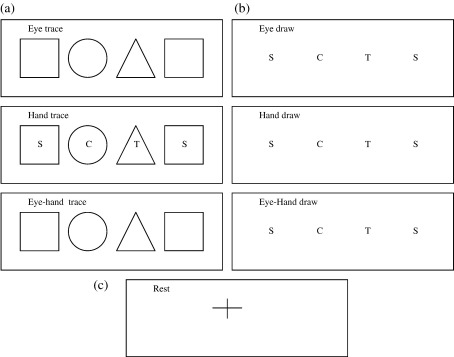
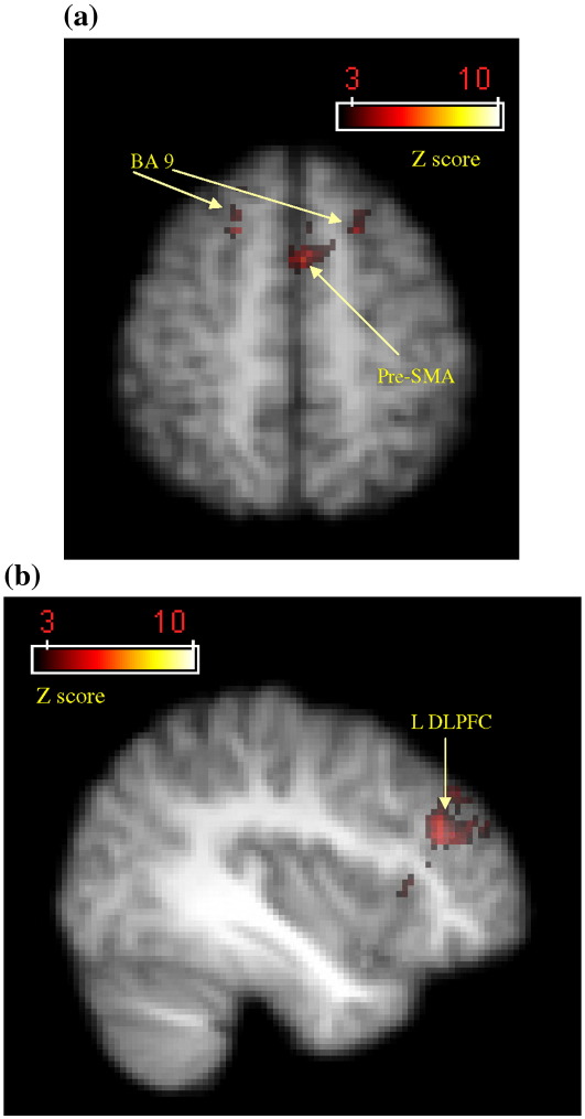
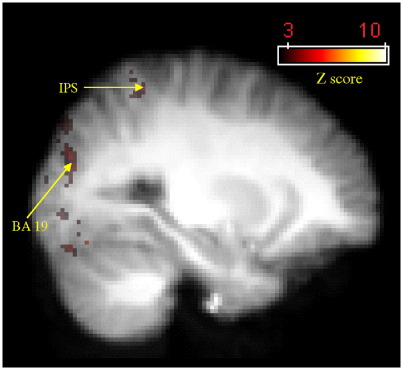
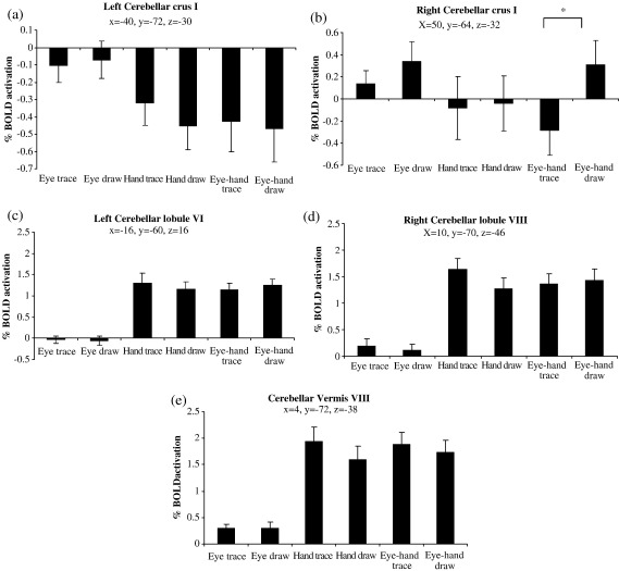
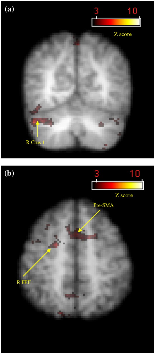
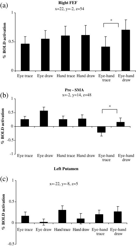
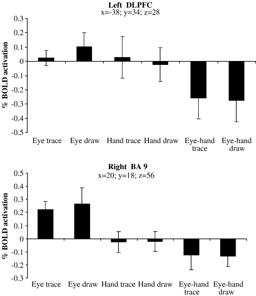
Externally cued movement is thought to preferentially involve cerebellar and premotor circuits whereas internally generated movement recruits basal ganglia, pre-supplementary motor cortex (pre-SMA) and dorsolateral prefrontal cortex (DLPFC). Tracing and drawing are exemplar externally and internally guided actions and Parkinson's patients and cerebellar patients show deficits in tracking and drawing, respectively. In this study we aimed to examine this external/internal distinction in healthy subjects using functional imaging. Ten healthy subjects performed tracing and drawing of simple geometric shapes using pencil and paper while in a 3-T fMRI scanner. Results indicated that compared to tracing, drawing generated greater activation in the right cerebellar crus I, bilateral pre-SMA, right dorsal premotor cortex and right frontal eye field. Tracing did not recruit any additional activation compared to drawing except in striate and extrastriate visual areas. Therefore, drawing recruited areas more frequently associated with cognitively challenging tasks, attention and memory, but basal ganglia and cerebellar activity did not differentiate tracing from drawing in the hypothesised manner. As our paradigm was of a simple, repetitive and static design, these results suggest that the task familiarity and the temporal nature of visual feedback in tracking tasks, compared to tracing, may be important contributing factors towards the degree of cerebellar involvement. Future studies comparing dynamic with static external cues and visual feedback may clarify the role of the cerebellum and basal ganglia in the visual guidance of drawing actions.
外部提示的运动被认为优先涉及小脑和运动前区回路,而内部产生的运动则需要基底神经节、补充运动前皮质(pre-SMA)和背外侧前额叶皮质(DLPFC)的参与。追踪和绘画分别是外部和内部引导动作的典型例子,帕金森病患者和小脑患者在追踪和绘画方面分别表现出缺陷。在本研究中,我们旨在使用功能成像检查健康受试者中的这种外部/内部差异。10名健康受试者在3-T功能磁共振成像扫描仪中使用铅笔和纸对简单几何形状进行追踪和绘画。结果表明,与追踪相比,绘画在右侧小脑脚I、双侧补充运动前皮质、右侧背侧运动前皮质和右侧额叶眼区产生了更大的激活。与绘画相比,追踪除了在纹状和纹外视觉区域外,没有引起任何额外的激活。因此,绘画激活的区域更常与认知挑战性任务、注意力和记忆相关,但基底神经节和小脑的活动并没有以假设的方式区分追踪和绘画。由于我们的范式是简单、重复和静态的设计,这些结果表明,与追踪相比,追踪任务中任务的熟悉程度和视觉反馈的时间性质可能是小脑参与程度的重要影响因素。未来比较动态与静态外部提示和视觉反馈的研究可能会阐明小脑和基底神经节在绘画动作视觉引导中的作用。