Xu Zheng, Choudhary Shilpa, Okada Yosuke, Voznesensky Olga, Alander Cynthia, Raisz Lawrence, Pilbeam Carol
Department of Medicine, Room N4051, MARB, MC5456, University of Connecticut Health Center, Farmington, 263 Farmington Avenue, Farmington, CT 06030, USA.
Bone. 2007 Jul;41(1):68-76. doi: 10.1016/j.bone.2007.03.009. Epub 2007 Mar 21.
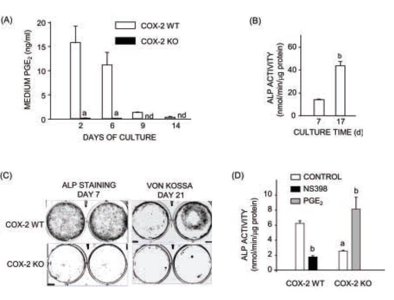
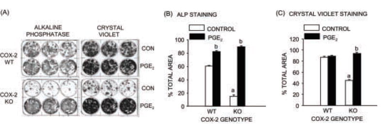
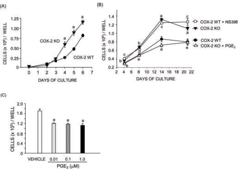
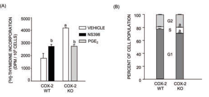
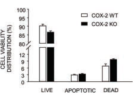
Cyclooxygenase-2 (COX-2) is highly expressed in osteoblasts, and COX-2 produced prostaglandins (PGs) can increase osteoblastic differentiation in vitro. The goal of this study was to examine effects of COX-2 expression on calvarial osteoblastic proliferation and apoptosis. Primary osteoblasts (POBs) were cultured from calvariae of COX-2 wild-type (WT) and knockout (KO) mice. POB proliferation was evaluated by (3)H-thymidine incorporation and analysis of cell replication and cell cycle distribution by flow cytometry. POB apoptosis was evaluated by annexin and PI staining on flow cytometry. As expected, PGE(2) production and alkaline phosphatase (ALP) activity were increased in WT cultures compared to KO cultures. In contrast, cell numbers were decreased in WT compared to KO cells by day 4 of culture. Proliferation, measured on days 3-7 of culture, was 2-fold greater in KO than in WT POBs and associated with decreased Go/G1 and increased S cell cycle distribution. There was no significant effect of COX-2 genotype on apoptosis under basal culture conditions on day 5 of culture. Cell growth was decreased in KO POBs by the addition of PGE(2) or a protein kinase A agonist and increased in WT POBs by the addition of NS398, a selective COX-2 inhibitor. In contrast, differentiation and cell growth in marrow stromal cell (MSC) cultures, evaluated by ALP and crystal violet staining respectively, were increased in MSCs from WT mice compared to MSCs from KO mice, and exogenous PGE(2) increased cell growth in KO MSC cultures. We conclude that PGs secondary to COX-2 expression decrease osteoblastic proliferation in cultured calvarial cells but increase growth of osteoblastic precursors in MSC cultures.
环氧化酶-2(COX-2)在成骨细胞中高表达,且COX-2产生的前列腺素(PGs)可在体外增加成骨细胞分化。本研究的目的是检测COX-2表达对颅骨成骨细胞增殖和凋亡的影响。从COX-2野生型(WT)和敲除(KO)小鼠的颅骨中培养原代成骨细胞(POBs)。通过³H-胸腺嘧啶核苷掺入以及流式细胞术分析细胞复制和细胞周期分布来评估POB增殖。通过流式细胞术的膜联蛋白和碘化丙啶染色评估POB凋亡。正如预期的那样,与KO培养物相比,WT培养物中PGE₂的产生和碱性磷酸酶(ALP)活性增加。相反,培养至第4天时,WT细胞中的细胞数量比KO细胞减少。在培养的第3 - 7天测量的增殖,KO的POB比WT的POB高2倍,且与Go/G1期减少和S期细胞周期分布增加相关。在培养第5天的基础培养条件下,COX-2基因型对凋亡没有显著影响。添加PGE₂或蛋白激酶A激动剂会使KO的POB细胞生长减少,而添加选择性COX-2抑制剂NS398会使WT的POB细胞生长增加。相反,分别通过ALP和结晶紫染色评估,WT小鼠的骨髓间充质细胞(MSC)培养物中的分化和细胞生长比KO小鼠的MSC培养物增加,且外源性PGE₂增加了KO的MSC培养物中的细胞生长。我们得出结论,COX-2表达产生的PGs会降低培养的颅骨细胞中的成骨细胞增殖,但会增加MSC培养物中成骨细胞前体的生长。