Rymer Jodi, Choh Vivian, Bharadwaj Shrikant, Padmanabhan Varuna, Modilevsky Laura, Jovanovich Elizabeth, Yeh Brenda, Zhang Zhan, Guan Huanxian, Payne W, Wildsoet Christine F
Wildsoet Lab, 588 Minor Hall, University of California-Berkeley, Berkeley CA 94720-2020, USA.
Exp Eye Res. 2007 Oct;85(4):431-42. doi: 10.1016/j.exer.2007.06.010. Epub 2007 Jun 21.
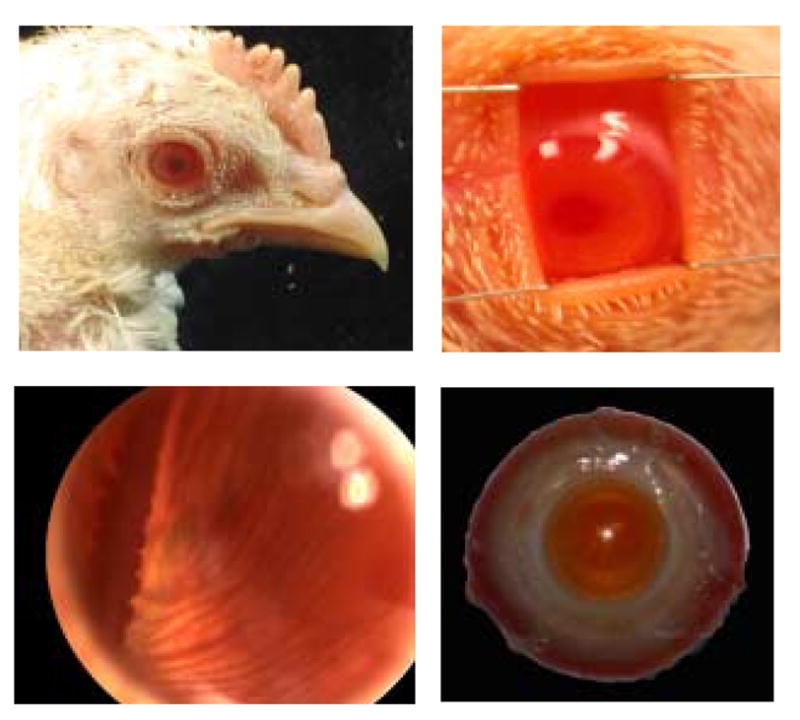
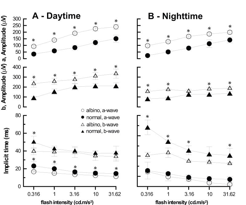
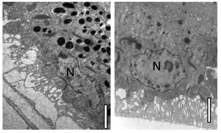
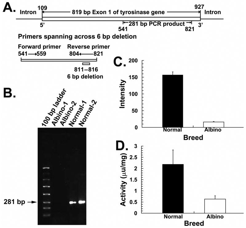
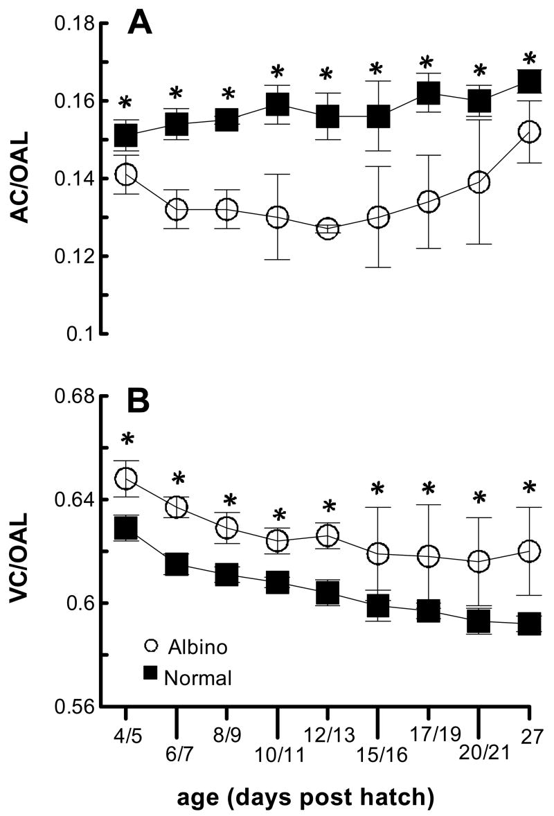
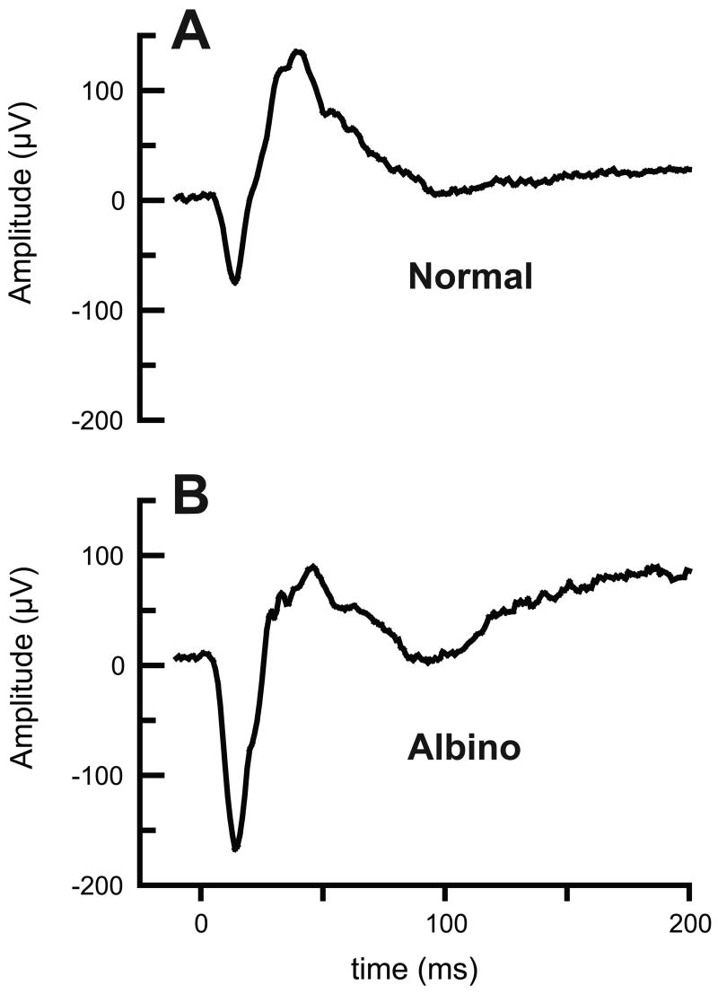
Albinism is associated with a variety of ocular anomalies including refractive errors. The purpose of this study was to investigate the ocular development of an albino chick line. The ocular development of both albino and normally pigmented chicks was monitored using retinoscopy to measure refractive errors and high frequency A-scan ultrasonography to measure axial ocular dimensions. Functional tests included an optokinetic nystagmus paradigm to assess visual acuity, and flash ERGs to assess retinal function. The underlying genetic abnormality was characterized using a gene microarray, PCR and a tyrosinase assay. The ultrastructure of the retinal pigment epithelium (RPE) was examined using transmission electron microscopy. PCR confirmed that the genetic abnormality in this line is a deletion in exon 1 of the tyrosinase gene. Tyrosinase gene expression in isolated RPE cells was minimally detectable, and there was minimal enzyme activity in albino feather bulbs. The albino chicks had pink eyes and their eyes transilluminated, reflecting the lack of melanin in all ocular tissues. All three main components, anterior chamber, crystalline lens and vitreous chamber, showed axial expansion over time in both normal and albino animals, but the anterior chambers of albino chicks were consistently shallower than those of normal chicks, while in contrast, their vitreous chambers were longer. Albino chicks remained relatively myopic, with higher astigmatism than the normally pigmented chicks, even though both groups underwent developmental emmetropization. Albino chicks had reduced visual acuity yet the ERG a- and b-wave components had larger amplitudes and shorter than normal implicit times. Developmental emmetropization occurs in the albino chick but is impaired, likely because of functional abnormalities in the RPE and/or retina as well as optical factors. In very young chicks the underlying genetic mutation may also contribute to refractive error and eye shape abnormalities.
白化病与包括屈光不正在内的多种眼部异常有关。本研究的目的是调查一种白化病雏鸡品系的眼部发育情况。使用检影法测量屈光不正,并使用高频A扫描超声检查测量眼轴尺寸,对白化病雏鸡和正常色素沉着雏鸡的眼部发育进行监测。功能测试包括使用视动性眼球震颤范式评估视力,以及使用闪光视网膜电图评估视网膜功能。使用基因微阵列、聚合酶链反应(PCR)和酪氨酸酶测定法对潜在的基因异常进行表征。使用透射电子显微镜检查视网膜色素上皮(RPE)的超微结构。PCR证实该品系的基因异常是酪氨酸酶基因外显子1的缺失。在分离的RPE细胞中,酪氨酸酶基因表达几乎检测不到,在白化病羽毛球中酶活性也极低。白化病雏鸡眼睛呈粉红色且可透照,这反映出所有眼部组织中都缺乏黑色素。前房、晶状体和玻璃体腔这三个主要成分在正常和白化病动物中均随时间呈现轴向扩张,但白化病雏鸡的前房始终比正常雏鸡的浅,而相比之下,它们的玻璃体腔更长。白化病雏鸡相对近视,散光比正常色素沉着的雏鸡更高,尽管两组都经历了发育性正视化过程。白化病雏鸡的视力下降,但其视网膜电图的a波和b波成分振幅更大,潜伏期比正常的短。白化病雏鸡会发生发育性正视化,但受到损害,这可能是由于RPE和/或视网膜的功能异常以及光学因素所致。在非常年幼的雏鸡中,潜在的基因突变也可能导致屈光不正和眼形异常。