Ho Beng-Choon
Department of Psychiatry, University of Iowa Carver College of Medicine, Iowa City, Iowa 52242, USA.
Schizophr Res. 2007 Nov;96(1-3):1-13. doi: 10.1016/j.schres.2007.08.001. Epub 2007 Aug 29.
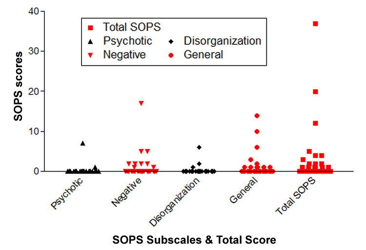
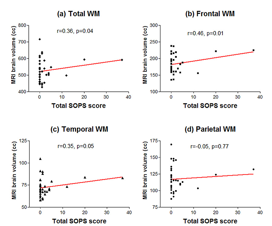
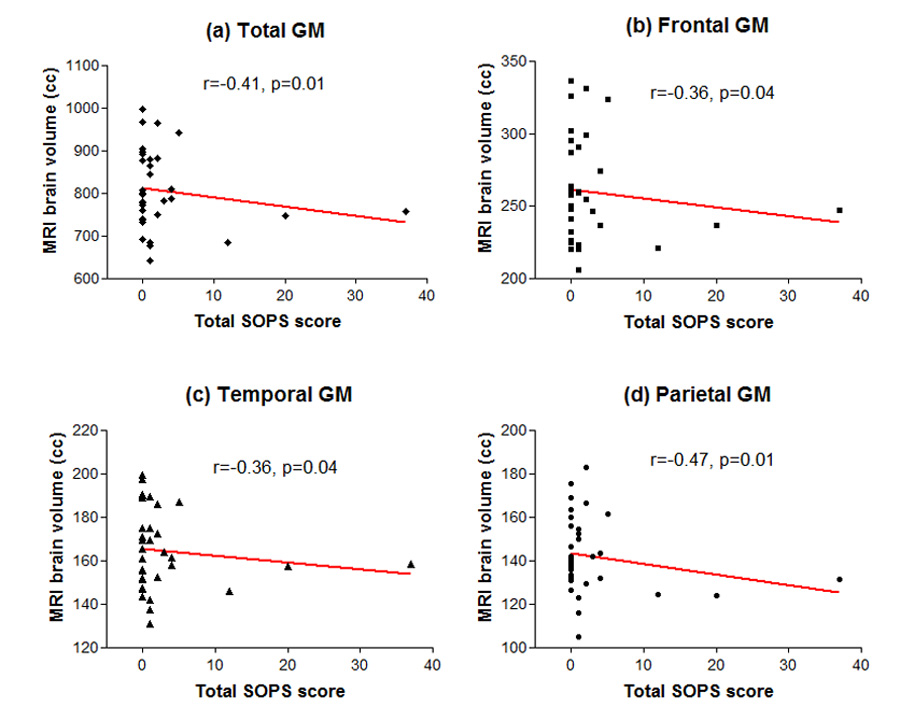
Schizophrenia is characterized by subtle but well-replicated total and regional (frontal and temporal) brain tissue volume deficits. Studies of individuals at-risk for developing schizophrenia suggest that the onset of brain volume decrement may closely pre-date overt manifestations of schizophrenia, making brain volume abnormalities potential predictors for early identification. In an ongoing longitudinal morphometric MRI study of young, nonpsychotic first- or second-degree relatives of schizophrenia probands, we compared brain volumes in 46 relatives who are still within age range for developing schizophrenia against comparison groups of 46 schizophrenia patients and 46 healthy volunteers without family history of schizophrenia. Relatives had similar brain volume abnormalities as schizophrenia patients albeit less severe. Relatives had significantly larger whole brain, frontal, temporal and parietal gray matter (GM) volumes than patients. Relatives also had significantly smaller frontal GM volumes than healthy volunteers. Both relatives and patients had significantly larger whole brain WM (specifically parietal WM) volumes compared to healthy volunteers. Abnormally greater WM volumes in relatives and patients are suggestive of genetically-mediated dysmaturation of the age-expected myelination during adolescence through mid adulthood. On prodromal symptoms assessed in relatives one year after MRI brain scans, initial GM deficits as well as larger WM volumes correlated significantly with greater severity of subsequent prodromal symptoms. Together with previous genetic high-risk studies of adolescent or young adult relatives, these findings indicate that premorbid MRI brain abnormalities may be of predictive value for the early identification of schizophrenia.
精神分裂症的特征是存在细微但已被充分证实的全脑及局部(额叶和颞叶)脑组织体积减少。对有患精神分裂症风险个体的研究表明,脑体积减少的起始可能在精神分裂症明显症状出现之前不久,这使得脑体积异常成为早期识别的潜在预测指标。在一项正在进行的针对精神分裂症先证者的年轻、无精神病的一级或二级亲属的纵向形态学MRI研究中,我们将46名仍处于患精神分裂症年龄范围内的亲属的脑体积与46名精神分裂症患者及46名无精神分裂症家族史的健康志愿者的对照组进行了比较。亲属与精神分裂症患者有相似的脑体积异常,尽管程度较轻。亲属的全脑、额叶、颞叶和顶叶灰质(GM)体积显著大于患者。亲属的额叶GM体积也显著小于健康志愿者。与健康志愿者相比,亲属和患者的全脑白质(特别是顶叶白质)体积均显著更大。亲属和患者异常增大的白质体积提示在青春期至成年中期,年龄预期的髓鞘形成存在基因介导的发育异常。在MRI脑部扫描一年后对亲属评估的前驱症状方面,最初的GM减少以及更大的WM体积与随后前驱症状的更严重程度显著相关。连同之前对青少年或年轻成年亲属的遗传高风险研究,这些发现表明病前MRI脑异常可能对精神分裂症的早期识别具有预测价值。