Kasai Kiyoto, Yamasue Hidenori, Gilbertson Mark W, Shenton Martha E, Rauch Scott L, Pitman Roger K
Department of Neuropsychiatry, Graduate School of Medicine, University of Tokyo, Tokyo, Japan.
Biol Psychiatry. 2008 Mar 15;63(6):550-6. doi: 10.1016/j.biopsych.2007.06.022. Epub 2007 Sep 7.
Controversy exists over the nature and origin of reduced regional brain volumes in posttraumatic stress disorder (PTSD). At issue is whether these reductions represent preexisting vulnerability factors for developing PTSD upon traumatic exposure or acquired PTSD signs due to the traumatic stress that caused the PTSD or the chronic stress of having the disorder (or both). We employed a case-control design in monozygotic twin pairs discordant for combat exposure to address the preexisting versus acquired origin of brain morphometric abnormalities in PTSD.
We used voxel-based morphometry to search for gray matter density reductions in magnetic resonance imaging (MRI) data obtained in a previous study of combat-exposed Vietnam veteran twins with (n = 18) versus without (n = 23) PTSD and their "high-risk" versus "low-risk" (respectively) identical combat-unexposed cotwins.
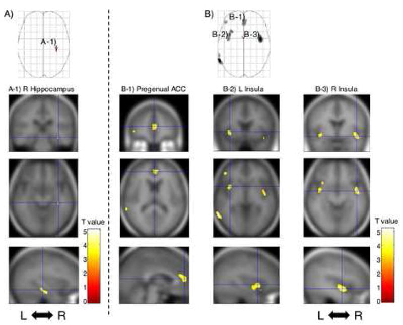
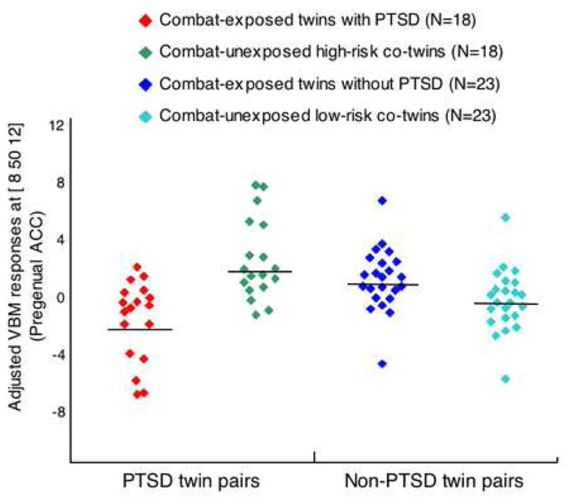
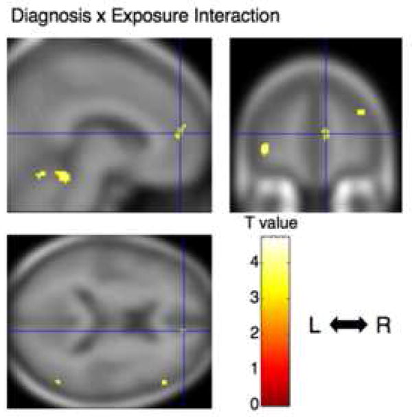
Compared with the combat-exposed twins without PTSD, the combat-exposed twins with PTSD showed significant gray matter density reductions in four predicted brain regions: right hippocampus, pregenual anterior cingulate cortex (ACC), and left and right insulae. There was a significant PTSD Diagnosis x Combat Exposure interaction in pregenual ACC in which combat-exposed PTSD twins had lower gray matter density than their own combat-unexposed cotwins as well as than the combat-exposed twins without PTSD and their cotwins.
The results point to gray matter volume diminutions in limbic and paralimbic structures in PTSD. The pattern of results obtained for pregenual ACC suggests that gray matter reduction in this region represents an acquired sign of PTSD consistent with stress-induced loss.
创伤后应激障碍(PTSD)患者局部脑容量减少的性质和起源存在争议。问题在于这些减少是代表创伤暴露时发生PTSD的预先存在的易患因素,还是由于导致PTSD的创伤应激或患有该疾病的慢性应激(或两者兼有)而获得的PTSD体征。我们采用病例对照设计,研究对象为单卵双胞胎中一方有战斗暴露经历,以探讨PTSD患者脑形态计量学异常是预先存在还是后天获得的。
我们使用基于体素的形态测量法,在先前一项针对有战斗暴露经历的越南退伍军人双胞胎的研究中获取的磁共振成像(MRI)数据中寻找灰质密度降低情况,这些双胞胎中,18例患有PTSD,23例未患PTSD,他们的同卵双胞胎分别为“高风险”和“低风险”(均无战斗暴露经历)。
与未患PTSD的有战斗暴露经历的双胞胎相比,患PTSD的有战斗暴露经历的双胞胎在四个预测脑区显示出显著的灰质密度降低:右侧海马体、膝前扣带回皮质(ACC)以及左右脑岛。在膝前ACC区域存在显著的PTSD诊断×战斗暴露交互作用,即有战斗暴露经历的PTSD双胞胎的灰质密度低于其未经历战斗的同卵双胞胎,也低于未患PTSD的有战斗暴露经历的双胞胎及其同卵双胞胎。
结果表明PTSD患者边缘和边缘旁结构的灰质体积减小。膝前ACC区域获得的结果模式表明,该区域灰质减少是PTSD的后天体征,与应激诱导的损失一致。