Bremner J Douglas
Department of Psychiatry and Behavioral Sciences, Emory University School of Medicine, Atlanta, Ga 30306, USA.
Dialogues Clin Neurosci. 2006;8(4):445-61. doi: 10.31887/DCNS.2006.8.4/jbremner.
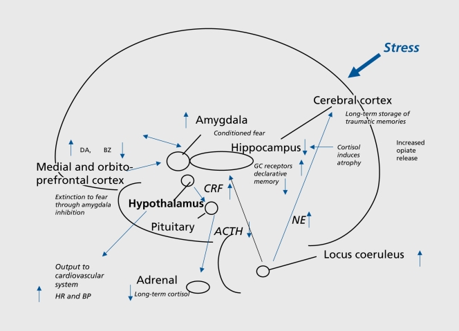
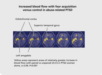
Brain areas implicated in the stress response include the amygdala, hippocampus, and prefrontal cortex. Traumatic stress can be associated with lasting changes in these brain areas. Traumatic stress is associated with increased cortisol and norepinephrine responses to subsequent stressors. Antidepressants have effects on the hippocampus that counteract the effects of stress. Findings from animal studies have been extended to patients with post-traumatic stress disorder (PTSD) showing smaller hippocampal and anterior cingulate volumes, increased amygdala function, and decreased medial prefrontal/anterior cingulate function. In addition, patients with PTSD show increased cortisol and norepinephrine responses to stress. Treatments that are efficacious for PTSD show a promotion of neurogenesis in animal studies, as well as promotion of memory and increased hippocampal volume in PTSD.
与应激反应相关的脑区包括杏仁核、海马体和前额叶皮质。创伤性应激可能与这些脑区的持久变化有关。创伤性应激与对后续应激源的皮质醇和去甲肾上腺素反应增加有关。抗抑郁药对海马体有作用,可抵消应激的影响。动物研究的结果已扩展到创伤后应激障碍(PTSD)患者,这些患者表现出海马体和前扣带回体积较小、杏仁核功能增强以及内侧前额叶/前扣带回功能降低。此外,PTSD患者对应激的皮质醇和去甲肾上腺素反应增加。在动物研究中,对PTSD有效的治疗方法显示出促进神经发生,以及在PTSD患者中促进记忆和增加海马体体积。