Zhang Xunli, Yin Huabing, Cooper Jon M, Haswell Stephen J
Department of Chemistry, The University of Hull, Hull, HU6 7RX, UK.
Anal Bioanal Chem. 2008 Feb;390(3):833-40. doi: 10.1007/s00216-007-1564-9. Epub 2007 Sep 12.
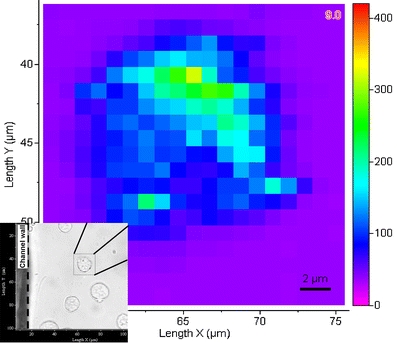
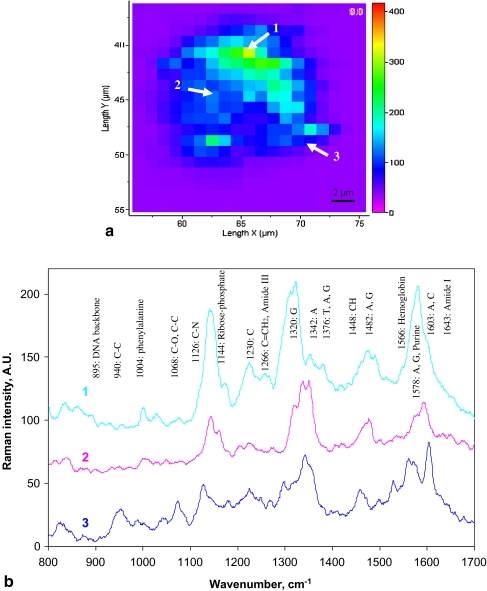
The integration of a range of technologies including microfluidics, surface-enhanced Raman scattering and confocal microspectroscopy has been successfully used to characterize in situ single living CHO (Chinese hamster ovary) cells with a high degree of spatial (in three dimensions) and temporal (1 s per spectrum) resolution. Following the introduction of a continuous flow of ionomycin, the real time spectral response from the cell was monitored during the agonist-evoked Ca(2+) flux process. The methodology described has the potential to be used for the study of the cellular dynamics of a range of signalling processes.
包括微流体技术、表面增强拉曼散射和共聚焦显微光谱学在内的一系列技术的整合已成功用于原位表征单个活的中国仓鼠卵巢(CHO)细胞,具有高度的空间(三维)和时间(每秒一个光谱)分辨率。在引入离子霉素的连续流后,在激动剂诱发的Ca(2+)通量过程中监测细胞的实时光谱响应。所描述的方法有潜力用于研究一系列信号传导过程的细胞动力学。