Knott V, Lapierre Y D, Fraser G, Johnson N
Institute of Mental Health Research/Department of Psychiatry and Psychology, University of Ottawa, Ontario.
J Psychiatry Neurosci. 1991 Nov;16(4):215-20.
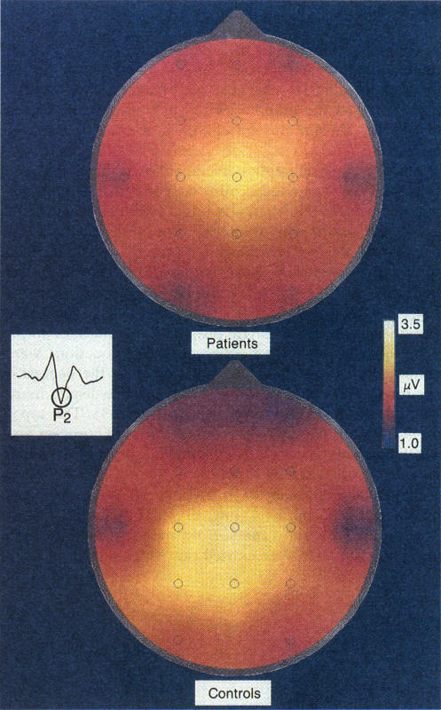
Neuroimaging studies of behavioral-induced anxiety in non-patients and of lactate-induced anxiety in panic disorder patients have indicated that normal and pathological anxiety may share a common pathway involving the temporal poles. As panic-related anxiety may reflect faulty temporopolar evaluative processing of input, the objective of this study was to examine sensory reactivity in panic disorder patients via scalp recordings of the late auditory evoked 'vertex' potential (LAEP) which appears to have a predominantly temporal lobe origin. Twelve patients diagnosed according to DSM-III criteria as panic disorder and ten normal controls served as subjects in this study. EEG was recorded from 16 scalp sites using a monopolar fronto-occipital derivation and LAEPs were separately averaged in response to four acoustic intensities. Analysis focused on group and electrode-site differences in the negative (N1) and positive (P2) component amplitudes of the LAEPs. Panic disorder patients were found to exhibit significantly larger N1 amplitudes across all stimulus intensities and across all recording sites. No significant group differences were observed with P2. Although the results provide indirect support for a temporal focus, other modulating influences must be considered in data interpretation.
针对非患者行为诱导性焦虑以及惊恐障碍患者乳酸诱导性焦虑的神经影像学研究表明,正常焦虑和病理性焦虑可能共享一条涉及颞极的共同通路。由于与惊恐相关的焦虑可能反映了对输入的颞极评估处理有误,本研究的目的是通过对晚期听觉诱发电位“头顶电位”(LAEP)进行头皮记录来检查惊恐障碍患者的感觉反应性,该电位似乎主要起源于颞叶。根据《精神疾病诊断与统计手册第三版》(DSM - III)标准诊断为惊恐障碍的12名患者和10名正常对照者作为本研究的受试者。使用单极额枕导联从16个头皮部位记录脑电图,并针对四种声音强度分别平均LAEP。分析集中在LAEP的负性(N1)和正性(P2)成分波幅的组间和电极部位差异。结果发现,在所有刺激强度和所有记录部位,惊恐障碍患者的N1波幅显著更大。P2未观察到显著的组间差异。尽管结果为颞叶焦点提供了间接支持,但在数据解释中必须考虑其他调节影响。