• 文献检索
  • 文档翻译
  • 深度研究
  • 学术资讯
  • Suppr Zotero 插件Zotero 插件
  • 邀请有礼
  • 套餐&价格
  • 历史记录
应用&插件
Suppr Zotero 插件Zotero 插件浏览器插件Mac 客户端Windows 客户端微信小程序
定价
高级版会员购买积分包购买API积分包
服务
文献检索文档翻译深度研究API 文档MCP 服务
关于我们
关于 Suppr公司介绍联系我们用户协议隐私条款
关注我们

Suppr 超能文献

核心技术专利:CN118964589B侵权必究
粤ICP备2023148730 号-1Suppr @ 2026

文献检索

告别复杂PubMed语法,用中文像聊天一样搜索,搜遍4000万医学文献。AI智能推荐,让科研检索更轻松。

立即免费搜索

文件翻译

保留排版,准确专业,支持PDF/Word/PPT等文件格式,支持 12+语言互译。

免费翻译文档

深度研究

AI帮你快速写综述,25分钟生成高质量综述,智能提取关键信息,辅助科研写作。

立即免费体验

图像分割对SK-BR-3细胞高内涵筛选数据质量的影响。

Impact of image segmentation on high-content screening data quality for SK-BR-3 cells.

作者信息

Hill Andrew A, LaPan Peter, Li Yizheng, Haney Steve

机构信息

Department of Biological Technologies, Wyeth Research, 35 CambridgePark Drive, Cambridge, MA 02140, USA.

出版信息

BMC Bioinformatics. 2007 Sep 14;8:340. doi: 10.1186/1471-2105-8-340.

DOI:10.1186/1471-2105-8-340
PMID:17868449
原文链接:https://pmc.ncbi.nlm.nih.gov/articles/PMC2080643/
Abstract

BACKGROUND

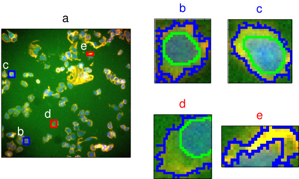
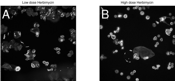
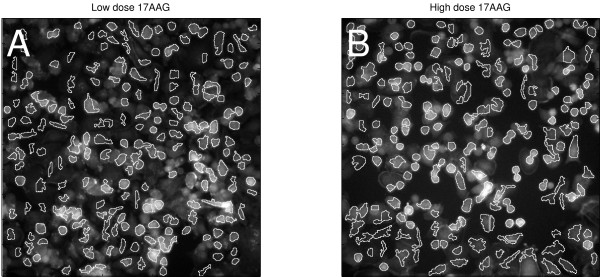
High content screening (HCS) is a powerful method for the exploration of cellular signalling and morphology that is rapidly being adopted in cancer research. HCS uses automated microscopy to collect images of cultured cells. The images are subjected to segmentation algorithms to identify cellular structures and quantitate their morphology, for hundreds to millions of individual cells. However, image analysis may be imperfect, especially for "HCS-unfriendly" cell lines whose morphology is not well handled by current image segmentation algorithms. We asked if segmentation errors were common for a clinically relevant cell line, if such errors had measurable effects on the data, and if HCS data could be improved by automated identification of well-segmented cells.

RESULTS

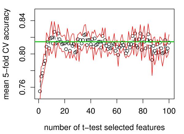
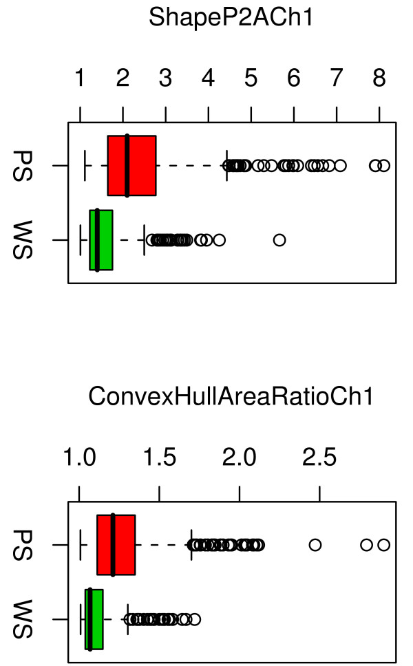
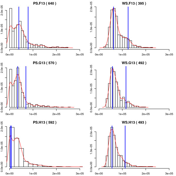
Cases of poor cell body segmentation occurred frequently for the SK-BR-3 cell line. We trained classifiers to identify SK-BR-3 cells that were well segmented. On an independent test set created by human review of cell images, our optimal support-vector machine classifier identified well-segmented cells with 81% accuracy. The dose responses of morphological features were measurably different in well- and poorly-segmented populations. Elimination of the poorly-segmented cell population increased the purity of DNA content distributions, while appropriately retaining biological heterogeneity, and simultaneously increasing our ability to resolve specific morphological changes in perturbed cells.

CONCLUSION

Image segmentation has a measurable impact on HCS data. The application of a multivariate shape-based filter to identify well-segmented cells improved HCS data quality for an HCS-unfriendly cell line, and could be a valuable post-processing step for some HCS datasets.

摘要

背景

高内涵筛选(HCS)是一种用于探索细胞信号传导和形态学的强大方法,正在癌症研究中迅速得到应用。HCS利用自动显微镜收集培养细胞的图像。这些图像经过分割算法处理,以识别细胞结构并对其形态进行定量分析,适用于数百到数百万个单个细胞。然而,图像分析可能并不完美,特别是对于那些形态不能被当前图像分割算法很好处理的“对HCS不友好”的细胞系。我们探讨了对于一个临床相关细胞系而言,分割错误是否常见,此类错误对数据是否有可测量的影响,以及通过自动识别分割良好的细胞能否改善HCS数据。

结果

SK-BR-3细胞系经常出现细胞体分割不佳的情况。我们训练分类器来识别分割良好的SK-BR-3细胞。在通过人工检查细胞图像创建的独立测试集上,我们的最优支持向量机分类器识别分割良好细胞的准确率为81%。在分割良好和不佳的细胞群体中,形态特征的剂量反应存在显著差异。去除分割不佳的细胞群体提高了DNA含量分布的纯度,同时适当保留了生物学异质性,并增强了我们解析受干扰细胞中特定形态变化的能力。

结论

图像分割对HCS数据有可测量的影响。应用基于多元形状的滤波器来识别分割良好的细胞,提高了对HCS不友好细胞系的HCS数据质量,对于一些HCS数据集而言,这可能是一个有价值的后处理步骤。

https://cdn.ncbi.nlm.nih.gov/pmc/blobs/02fe/2080643/1767905e90d3/1471-2105-8-340-9.jpg
https://cdn.ncbi.nlm.nih.gov/pmc/blobs/02fe/2080643/abe2030edfcc/1471-2105-8-340-1.jpg
https://cdn.ncbi.nlm.nih.gov/pmc/blobs/02fe/2080643/63000f651271/1471-2105-8-340-2.jpg
https://cdn.ncbi.nlm.nih.gov/pmc/blobs/02fe/2080643/043d505df6d6/1471-2105-8-340-3.jpg
https://cdn.ncbi.nlm.nih.gov/pmc/blobs/02fe/2080643/8303f8d426b1/1471-2105-8-340-4.jpg
https://cdn.ncbi.nlm.nih.gov/pmc/blobs/02fe/2080643/94183a82d70a/1471-2105-8-340-5.jpg
https://cdn.ncbi.nlm.nih.gov/pmc/blobs/02fe/2080643/04af1b5d0926/1471-2105-8-340-6.jpg
https://cdn.ncbi.nlm.nih.gov/pmc/blobs/02fe/2080643/d74406f47d5c/1471-2105-8-340-7.jpg
https://cdn.ncbi.nlm.nih.gov/pmc/blobs/02fe/2080643/bbc0b06cd8af/1471-2105-8-340-8.jpg
https://cdn.ncbi.nlm.nih.gov/pmc/blobs/02fe/2080643/1767905e90d3/1471-2105-8-340-9.jpg

相似文献

1
Impact of image segmentation on high-content screening data quality for SK-BR-3 cells.图像分割对SK-BR-3细胞高内涵筛选数据质量的影响。
BMC Bioinformatics. 2007 Sep 14;8:340. doi: 10.1186/1471-2105-8-340.
2
An automated feedback system with the hybrid model of scoring and classification for solving over-segmentation problems in RNAi high content screening.一种具有评分和分类混合模型的自动反馈系统,用于解决RNA干扰高内涵筛选中的过分割问题。
J Microsc. 2007 May;226(Pt 2):121-32. doi: 10.1111/j.1365-2818.2007.01762.x.
3
A computerized cellular imaging system for high content analysis in Monastrol suppressor screens.用于Monastrol抑制剂筛选中高内涵分析的计算机化细胞成像系统。
J Biomed Inform. 2006 Apr;39(2):115-25. doi: 10.1016/j.jbi.2005.05.008. Epub 2005 Jun 22.
4
A fast, fully automated cell segmentation algorithm for high-throughput and high-content screening.一种用于高通量和高内涵筛选的快速、全自动细胞分割算法。
Cytometry A. 2008 Oct;73(10):958-64. doi: 10.1002/cyto.a.20627.
5
Analysis and recognition of touching cell images based on morphological structures.基于形态结构的接触细胞图像分析与识别
Comput Biol Med. 2009 Jan;39(1):27-39. doi: 10.1016/j.compbiomed.2008.10.006. Epub 2008 Dec 12.
6
Template-driven segmentation of confocal microscopy images.共聚焦显微镜图像的模板驱动分割
Comput Methods Programs Biomed. 2008 Mar;89(3):239-47. doi: 10.1016/j.cmpb.2007.11.007.
7
Automated Arabidopsis plant root cell segmentation based on SVM classification and region merging.基于支持向量机分类和区域合并的拟南芥植物根细胞自动分割
Comput Biol Med. 2009 Sep;39(9):785-93. doi: 10.1016/j.compbiomed.2009.06.008. Epub 2009 Jul 14.
8
A multidimensional segmentation evaluation for medical image data.医学图像数据的多维度分割评估
Comput Methods Programs Biomed. 2009 Nov;96(2):108-24. doi: 10.1016/j.cmpb.2009.04.009. Epub 2009 May 14.
9
Cell nuclei and cytoplasm joint segmentation using the sliding band filter.使用滑动带滤波器进行细胞核和细胞质联合分割。
IEEE Trans Med Imaging. 2010 Aug;29(8):1463-73. doi: 10.1109/TMI.2010.2048253. Epub 2010 Jun 3.
10
Performance evaluation and benchmarking of six-page segmentation algorithms.六种页面分割算法的性能评估与基准测试
IEEE Trans Pattern Anal Mach Intell. 2008 Jun;30(6):941-54. doi: 10.1109/TPAMI.2007.70837.

引用本文的文献

1
NeuronAlg: An Innovative Neuronal Computational Model for Immunofluorescence Image Segmentation.神经元算法:一种用于免疫荧光图像分割的创新神经元计算模型。
Sensors (Basel). 2023 May 9;23(10):4598. doi: 10.3390/s23104598.
2
A novel measure and significance testing in data analysis of cell image segmentation.细胞图像分割数据分析中的一种新测量方法及显著性检验
BMC Bioinformatics. 2017 Mar 14;18(1):168. doi: 10.1186/s12859-017-1527-x.
3
Semi-automated discrimination of retinal pigmented epithelial cells in two-photon fluorescence images of mouse retinas.

本文引用的文献

1
High content cellular screening.高内涵细胞筛选
Curr Opin Chem Biol. 2006 Aug;10(4):316-20. doi: 10.1016/j.cbpa.2006.06.004. Epub 2006 Jun 21.
2
Validating cancer drug targets.验证癌症药物靶点。
Nature. 2006 May 25;441(7092):451-6. doi: 10.1038/nature04873.
3
A computerized cellular imaging system for high content analysis in Monastrol suppressor screens.用于Monastrol抑制剂筛选中高内涵分析的计算机化细胞成像系统。
小鼠视网膜双光子荧光图像中视网膜色素上皮细胞的半自动识别
Biomed Opt Express. 2015 Jul 23;6(8):3032-52. doi: 10.1364/BOE.6.003032. eCollection 2015 Aug 1.
4
Ranked retrieval of segmented nuclei for objective assessment of cancer gene repositioning.分段核的分级检索,用于客观评估癌症基因重定位。
BMC Bioinformatics. 2012 Sep 12;13:232. doi: 10.1186/1471-2105-13-232.
5
Automatic segmentation and supervised learning-based selection of nuclei in cancer tissue images.自动分割和基于监督学习的癌症组织图像细胞核选择。
Cytometry A. 2012 Sep;81(9):743-54. doi: 10.1002/cyto.a.22097. Epub 2012 Jul 31.
6
Gebiss: an ImageJ plugin for the specification of ground truth and the performance evaluation of 3D segmentation algorithms.牙列:一个用于指定真实情况和评估 3D 分割算法性能的 ImageJ 插件。
BMC Bioinformatics. 2011 Jun 13;12:232. doi: 10.1186/1471-2105-12-232.
7
Generating 'omic knowledge': the role of informatics in high content screening.生成“组学知识”:信息学在高内涵筛选中的作用。
Comb Chem High Throughput Screen. 2009 Nov;12(9):917-25. doi: 10.2174/138620709789383259.
J Biomed Inform. 2006 Apr;39(2):115-25. doi: 10.1016/j.jbi.2005.05.008. Epub 2005 Jun 22.
4
Genome-wide analysis of human kinases in clathrin- and caveolae/raft-mediated endocytosis.网格蛋白及小窝/脂筏介导的内吞作用中人类激酶的全基因组分析。
Nature. 2005 Jul 7;436(7047):78-86. doi: 10.1038/nature03571. Epub 2005 May 11.
5
An unbiased cell morphology-based screen for new, biologically active small molecules.一种基于细胞形态学的无偏差筛选新型生物活性小分子的方法。
PLoS Biol. 2005 May;3(5):e128. doi: 10.1371/journal.pbio.0030128. Epub 2005 Apr 5.
6
Multidimensional drug profiling by automated microscopy.通过自动显微镜进行多维药物分析
Science. 2004 Nov 12;306(5699):1194-8. doi: 10.1126/science.1100709.
7
Enhanced sensitization to taxol-induced apoptosis by herceptin pretreatment in ErbB2-overexpressing breast cancer cells.赫赛汀预处理增强ErbB2过表达乳腺癌细胞对紫杉醇诱导凋亡的敏感性。
Cancer Res. 2002 Oct 15;62(20):5703-10.
8
A new human antitumor immunoreagent specific for ErbB2.一种新的针对ErbB2的人源抗肿瘤免疫试剂。
Clin Cancer Res. 2002 Jun;8(6):1710-9.
9
Treatment of meningeal breast cancer xenografts in the rat using an anti-p185/HER2 antibody.使用抗p185/HER2抗体治疗大鼠脑膜乳腺癌异种移植瘤。
Clin Cancer Res. 2001 Jul;7(7):2050-6.