Fan Rong, DeFilippis Kelly, Van Nostrand William E
Department of Medicine, Stony Brook University, Stony Brook, NY 11794 USA.
J Neuroinflammation. 2007 Sep 18;4:22. doi: 10.1186/1742-2094-4-22.
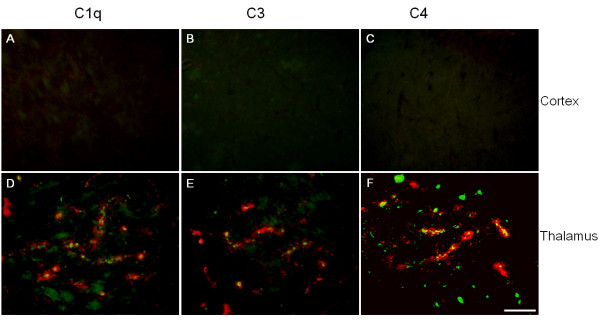
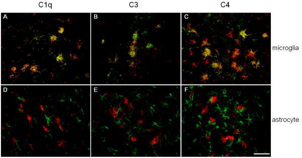
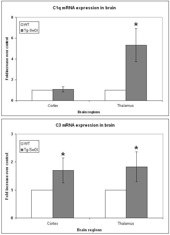
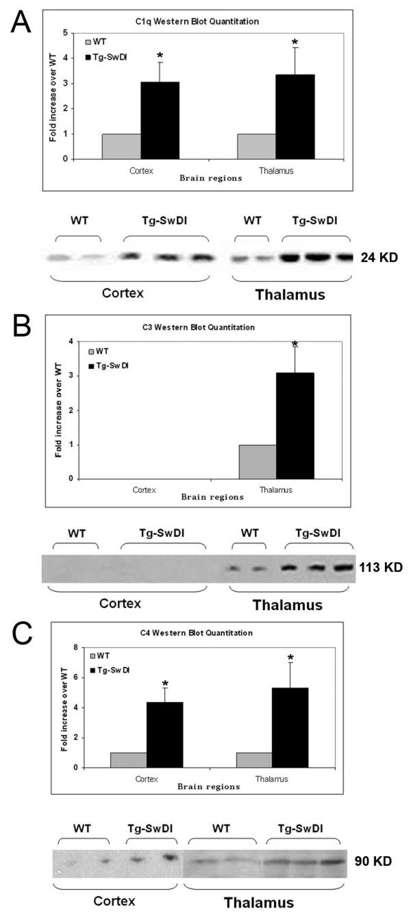
The deposition of amyloid beta-protein (A beta) in cerebral vasculature, known as cerebral amyloid angiopathy (CAA), is a common pathological feature of Alzheimer's disease and related disorders. In familial forms of CAA single mutations in the A beta peptide have been linked to the increase of vascular A beta deposits accompanied by a strong localized activation of glial cells and elevated expression of neuroinflammatory mediators including complement proteins. We have developed human amyloid-beta precursor protein transgenic mice harboring two CAA A beta mutations (Dutch E693Q and Iowa D694N) that mimic the prevalent cerebral microvascular A beta deposition observed in those patients, and the Swedish mutations (K670N/M671L) to increase A beta production. In these Tg-SwDI mice, we have reported predominant fibrillar A beta along microvessels in the thalamic region and diffuse plaques in cortical region. Concurrently, activated microglia and reactive astrocytes have been detected primarily in association with fibrillar cerebral microvascular A beta in this model. Here we show that three native complement components in classical and alternative complement pathways, C1q, C3, and C4, are elevated in Tg-SwDI mice in regions rich in fibrillar microvascular A beta. Immunohistochemical staining of all three proteins was increased in thalamus, hippocampus, and subiculum, but not frontal cortex. Western blot analysis showed significant increases of all three proteins in the thalamic region (with hippocampus) as well as the cortical region, except C3 that was below detection level in cortex. Also, in the thalamic region (with hippocampus), C1q and C3 mRNAs were significantly up-regulated. These complement proteins appeared to be expressed largely by activated microglial cells associated with the fibrillar microvascular A beta deposits. Our findings demonstrate that Tg-SwDI mice exhibit elevated complement protein expression in response to fibrillar vascular A beta deposition that is observed in patients with familial CAA.
淀粉样β蛋白(Aβ)在脑脉管系统中的沉积,即脑淀粉样血管病(CAA),是阿尔茨海默病及相关疾病的常见病理特征。在家族性CAA中,Aβ肽的单个突变与血管Aβ沉积物的增加有关,同时伴有胶质细胞的强烈局部激活以及包括补体蛋白在内的神经炎症介质表达升高。我们构建了携带两种CAA Aβ突变(荷兰人型E693Q和爱荷华型D694N)的人淀粉样β前体蛋白转基因小鼠,这些突变模拟了在那些患者中观察到的普遍的脑微血管Aβ沉积,以及瑞典突变(K670N/M671L)以增加Aβ的产生。在这些Tg-SwDI小鼠中,我们报道了丘脑区域微血管周围主要为纤维状Aβ以及皮质区域有弥漫性斑块。同时,在该模型中主要检测到活化的小胶质细胞和反应性星形胶质细胞与纤维状脑微血管Aβ相关。在此我们表明,经典和替代补体途径中的三种天然补体成分C1q、C3和C4在富含纤维状微血管Aβ的区域的Tg-SwDI小鼠中升高。这三种蛋白的免疫组织化学染色在丘脑、海马和下托中增加,但在额叶皮质中未增加。蛋白质印迹分析显示,除了皮质中C3低于检测水平外,丘脑区域(包括海马)以及皮质区域中所有三种蛋白均显著增加。此外,在丘脑区域(包括海马),C1q和C3的mRNA显著上调。这些补体蛋白似乎主要由与纤维状微血管Aβ沉积物相关的活化小胶质细胞表达。我们的研究结果表明,Tg-SwDI小鼠对家族性CAA患者中观察到的纤维状血管Aβ沉积有补体蛋白表达升高的反应。