Braun Rod D, Vistisen Kerry S
Department of Anatomy and Cell Biology, Wayne State University School of Medicine, 540 E. Canfield Avenue, Detroit, MI 48201, USA.
Invest Ophthalmol Vis Sci. 2008 Jan;49(1):16-22. doi: 10.1167/iovs.07-0379.
The purpose of this study was to test the hypothesis that the volume of primary orthotopic human choroidal melanoma xenografts can be quantified noninvasively in the same nude rat over time using a portable, high-resolution, high-frequency ultrasound (HF-US) system.
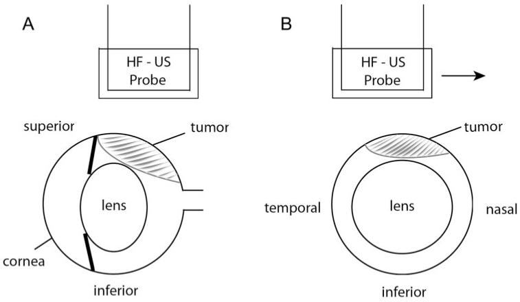
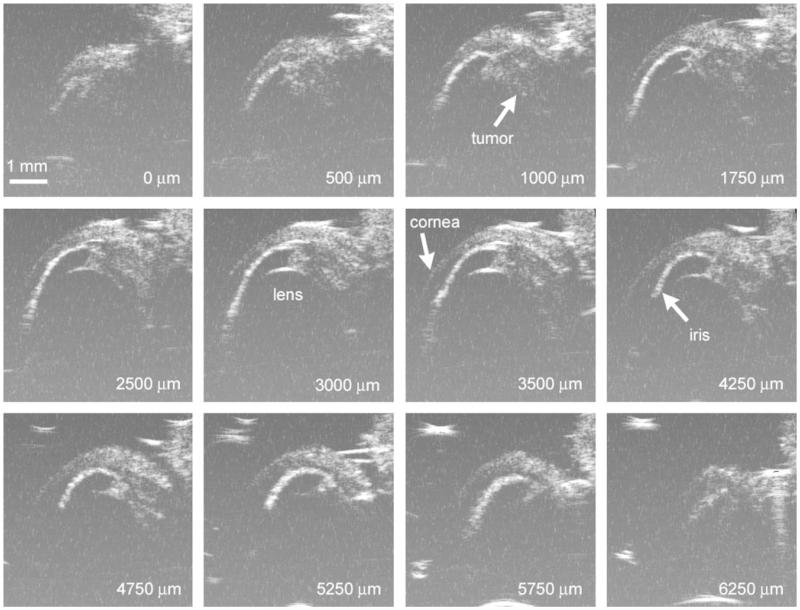
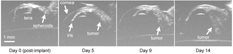
C918 human choroidal melanoma spheroids were implanted in the superior suprachoroidal space of 26 WAG/RijHsd-rnu nude rats. Fourteen rats were anesthetized 14 days after tumor implantation, and HF-US B-scan images of the tumor-bearing eye were captured at 250-mum intervals. Tumor areas were measured on each image and numerically integrated to calculate volume. Tumor volumes were also estimated from serial histologic sections in six rats. Twelve other rats were anesthetized and weighed every 4 to 5 days after implantation for 2 weeks, and HF-US B-scan image series were acquired for subsequent measurement of tumor volume.
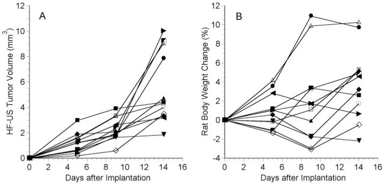
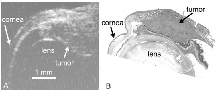
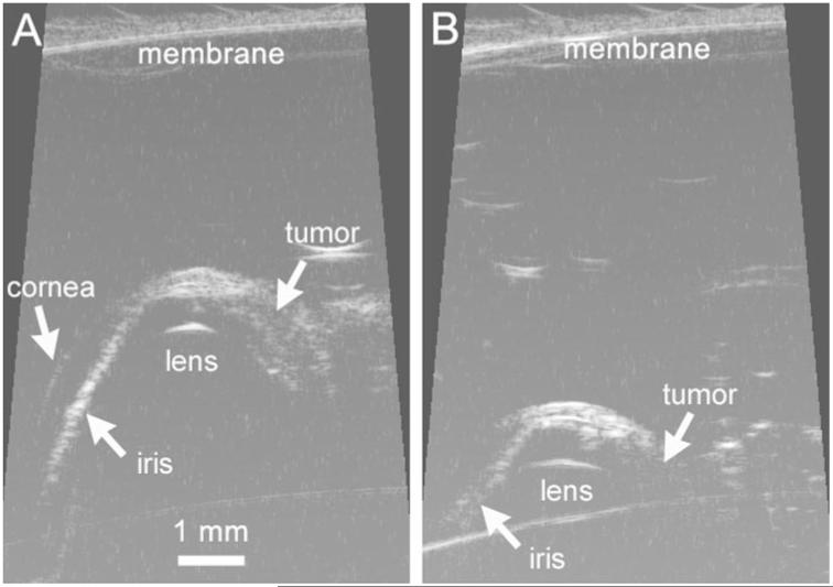
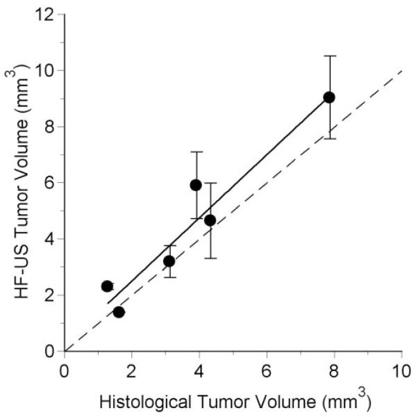
Tumors could be visualized as heterogeneous, relatively hyperechoic regions in the superior portion of the eye. These regions were verified as tumor by comparison with histologic sections, and histologic and HF-US volumes were highly correlated (r = 0.961; P = 0.002). For the determination of HF-US volume, the intraobserver variability was 9.7% +/- 5.1% (n = 8), and the coefficient of variation for multiple measurements was 12.1% +/- 6.8% (n = 12). Tumor volume could be repeatedly measured in the same rat every 4 to 5 days for 2 weeks without significant weight loss.
HF-US is a safe, practical method to measure tumor volume in the same nude rat over time in this orthotopic xenograft model of human choroidal melanoma.
本研究的目的是验证以下假设:使用便携式高分辨率高频超声(HF-US)系统,可在同一裸鼠体内随时间对原发性原位人脉络膜黑色素瘤异种移植瘤的体积进行无创定量。
将C918人脉络膜黑色素瘤球体植入26只WAG/RijHsd-rnu裸鼠的脉络膜上腔。14只大鼠在肿瘤植入后14天接受麻醉,以250μm的间隔获取荷瘤眼的HF-US B超图像。在每张图像上测量肿瘤面积,并进行数值积分以计算体积。还从6只大鼠的连续组织切片中估计肿瘤体积。另外12只大鼠在植入后每4至5天接受麻醉并称重,持续2周,获取HF-US B超图像系列以用于后续肿瘤体积测量。
肿瘤可在眼上部显示为不均匀的相对高回声区。通过与组织切片比较,这些区域被证实为肿瘤,组织学体积与HF-US体积高度相关(r = 0.961;P = 0.002)。对于HF-US体积的测定,观察者内变异性为9.7%±5.1%(n = 8),多次测量的变异系数为12.1%±6.8%(n = 12)。在2周内可每4至5天在同一只大鼠中重复测量肿瘤体积,且大鼠体重无明显减轻。
在这人脉络膜黑色素瘤原位异种移植模型中,HF-US是一种安全、实用的方法,可在同一裸鼠体内随时间测量肿瘤体积。