• 文献检索
  • 文档翻译
  • 深度研究
  • 学术资讯
  • Suppr Zotero 插件Zotero 插件
  • 邀请有礼
  • 套餐&价格
  • 历史记录
应用&插件
Suppr Zotero 插件Zotero 插件浏览器插件Mac 客户端Windows 客户端微信小程序
定价
高级版会员购买积分包购买API积分包
服务
文献检索文档翻译深度研究API 文档MCP 服务
关于我们
关于 Suppr公司介绍联系我们用户协议隐私条款
关注我们

Suppr 超能文献

核心技术专利:CN118964589B侵权必究
粤ICP备2023148730 号-1Suppr @ 2026

文献检索

告别复杂PubMed语法,用中文像聊天一样搜索,搜遍4000万医学文献。AI智能推荐,让科研检索更轻松。

立即免费搜索

文件翻译

保留排版,准确专业,支持PDF/Word/PPT等文件格式,支持 12+语言互译。

免费翻译文档

深度研究

AI帮你快速写综述,25分钟生成高质量综述,智能提取关键信息,辅助科研写作。

立即免费体验

使用9.4T反转恢复脉冲序列通过延迟钆增强磁共振成像评估小鼠心肌梗死情况。

Assessment of myocardial infarction in mice by late gadolinium enhancement MR imaging using an inversion recovery pulse sequence at 9.4T.

作者信息

Chapon Catherine, Herlihy Amy H, Bhakoo Kishore K

机构信息

Stem Cell Imaging, MRC Clinical Sciences Centre, Hammersmith Hospital, Imperial College London, UK.

出版信息

J Cardiovasc Magn Reson. 2008 Jan 24;10(1):6. doi: 10.1186/1532-429X-10-6.

DOI:10.1186/1532-429X-10-6
PMID:18272007
原文链接:https://pmc.ncbi.nlm.nih.gov/articles/PMC2244610/
Abstract

PURPOSE

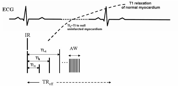
To demonstrate the feasibility of using an inversion recovery pulse sequence and to define the optimal inversion time (TI) to assess myocardial infarction in mice by late gadolinium enhancement (LGE) MRI at 9.4T, and to obtain the maximal contrast between the infarcted and the viable myocardium.

METHODS

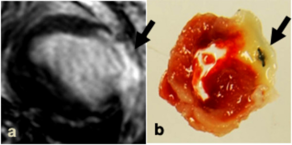
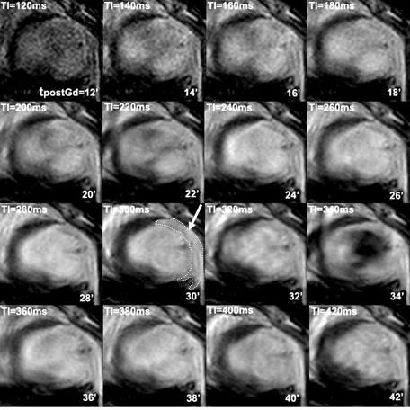
MRI was performed at 9.4T in mice, two days after induction of myocardial infarction (n = 4). For cardiovascular MR imaging, a segmented magnetization-prepared fast low angle shot (MP-FLASH) sequence was used with varied TIs ranging from 40 to 420 ms following administration of gadolinium-DTPA at 0.6 mmol/kg. Contrast-to-noise (CNR) and signal-to-noise ratio (SNR) were measured and compared for each myocardial region of interest (ROI).

RESULTS

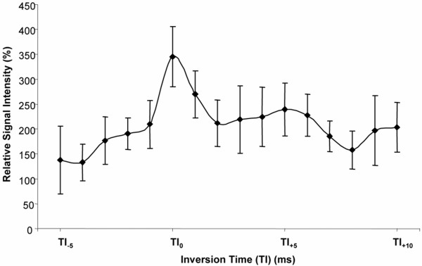
The optimal TI, which corresponded to a minimum SNR in the normal myocardium, was 268 ms +/- 27.3. The SNR in the viable myocardium was significantly different from that found in the infarcted myocardium (17.2 +/- 2.4 vs 82.1 +/- 10.8; p = 0.006) leading to a maximal relative SI (Signal Intensity) between those two areas (344.9 +/- 60.4).

CONCLUSION

Despite the rapid heart rate in mice, our study demonstrates that LGE MRI can be performed at 9.4T using a protocol similar to the one used for clinical MR diagnosis of myocardial infarction.

摘要

目的

通过9.4T场强下的延迟钆增强(LGE)磁共振成像(MRI),证明使用反转恢复脉冲序列评估小鼠心肌梗死的可行性,确定最佳反转时间(TI),并获得梗死心肌与存活心肌之间的最大对比度。

方法

在诱导心肌梗死后两天(n = 4),对小鼠进行9.4T场强的MRI检查。对于心血管磁共振成像,在注射0.6 mmol/kg钆喷酸葡胺后,使用分段磁化准备快速低角度激发(MP-FLASH)序列,TI范围为40至420 ms。测量并比较每个心肌感兴趣区域(ROI)的对比噪声比(CNR)和信噪比(SNR)。

结果

对应于正常心肌最小SNR的最佳TI为268 ms±27.3。存活心肌中的SNR与梗死心肌中的SNR有显著差异(17.2±2.4对82.1±10.8;p = 0.006),导致这两个区域之间的最大相对信号强度(SI)(344.9±60.4)。

结论

尽管小鼠心率较快,但我们的研究表明,使用与心肌梗死临床磁共振诊断类似的方案,可在9.4T场强下进行LGE MRI检查。

https://cdn.ncbi.nlm.nih.gov/pmc/blobs/2651/2244610/e335edb24cb7/1532-429X-10-6-4.jpg
https://cdn.ncbi.nlm.nih.gov/pmc/blobs/2651/2244610/852ce0ece073/1532-429X-10-6-1.jpg
https://cdn.ncbi.nlm.nih.gov/pmc/blobs/2651/2244610/8718accc5a61/1532-429X-10-6-2.jpg
https://cdn.ncbi.nlm.nih.gov/pmc/blobs/2651/2244610/27f215db62a3/1532-429X-10-6-3.jpg
https://cdn.ncbi.nlm.nih.gov/pmc/blobs/2651/2244610/e335edb24cb7/1532-429X-10-6-4.jpg

相似文献

1
Assessment of myocardial infarction in mice by late gadolinium enhancement MR imaging using an inversion recovery pulse sequence at 9.4T.使用9.4T反转恢复脉冲序列通过延迟钆增强磁共振成像评估小鼠心肌梗死情况。
J Cardiovasc Magn Reson. 2008 Jan 24;10(1):6. doi: 10.1186/1532-429X-10-6.
2
Phase-sensitive inversion-recovery MR imaging in the detection of myocardial infarction.用于检测心肌梗死的相敏反转恢复磁共振成像
Radiology. 2005 Dec;237(3):854-60. doi: 10.1148/radiol.2373041483.
3
Single-shot inversion recovery TrueFISP for assessment of myocardial infarction.用于评估心肌梗死的单次激发反转恢复快速成像稳态自由进动序列
AJR Am J Roentgenol. 2006 Mar;186(3):627-33. doi: 10.2214/AJR.04.0746.
4
Gadobutrol for magnetic resonance imaging of chronic myocardial infarction: intraindividual comparison with gadopentetate dimeglumine.钆布醇用于慢性心肌梗死磁共振成像:与马根维显的个体内比较。
Invest Radiol. 2012 Mar;47(3):183-8. doi: 10.1097/RLI.0b013e318236e354.
5
Ultra low-dose of gadobenate dimeglumine for late gadolinium enhancement (LGE) imaging in acute myocardial infarction: a feasibility study.超低剂量钆布醇用于急性心肌梗死延迟钆增强(LGE)成像的可行性研究
Eur J Radiol. 2014 Dec;83(12):2151-2158. doi: 10.1016/j.ejrad.2014.09.002. Epub 2014 Sep 28.
6
Comparison of fast multi-slice and standard segmented techniques for detection of late gadolinium enhancement in ischemic and non-ischemic cardiomyopathy - a prospective clinical cardiovascular magnetic resonance trial.比较快速多层面和标准分段技术在缺血性和非缺血性心肌病中的延迟钆增强检测 - 一项前瞻性临床心血管磁共振试验。
J Cardiovasc Magn Reson. 2018 Feb 19;20(1):13. doi: 10.1186/s12968-018-0434-2.
7
Single-dose gadobutrol in comparison with single-dose gadobenate dimeglumine for magnetic resonance imaging of chronic myocardial infarction at 3 T.3T 下单剂量钆布醇与单剂量钆贝葡胺用于慢性心肌梗死磁共振成像的比较
Invest Radiol. 2014 Nov;49(11):728-34. doi: 10.1097/RLI.0000000000000076.
8
Effect of inversion time on the precision of myocardial late gadolinium enhancement quantification evaluated with synthetic inversion recovery MR imaging.反转时间对采用合成反转恢复磁共振成像评估心肌延迟钆增强定量精度的影响。
Eur Radiol. 2017 Aug;27(8):3235-3243. doi: 10.1007/s00330-016-4665-z. Epub 2017 Jan 3.
9
Late gadolinium enhancement magnetic resonance imaging for the assessment of myocardial infarction: comparison of image quality between single and double doses of contrast agents.钆延迟强化磁共振成像用于评估心肌梗死:单剂量与双剂量造影剂的图像质量比较
Int J Cardiovasc Imaging. 2014 Dec;30 Suppl 2:129-35. doi: 10.1007/s10554-014-0505-x. Epub 2014 Aug 3.
10
Rapid assessment of myocardial infarct size in rodents using multi-slice inversion recovery late gadolinium enhancement CMR at 9.4T.在 9.4T 场强下使用多层面反转恢复晚期钆增强 CMR 快速评估啮齿类动物的心肌梗死面积。
J Cardiovasc Magn Reson. 2011 Sep 5;13(1):44. doi: 10.1186/1532-429X-13-44.

引用本文的文献

1
Left ventricle strain and T1 mapping evaluation in a mouse model with myocardial infarction.心肌梗死小鼠模型中的左心室应变和T1映射评估
Sci Rep. 2025 Aug 12;15(1):29579. doi: 10.1038/s41598-025-09699-0.
2
Late Gadolinium Enhancement of Nonischemic Cardiomyopathy at 5.0 T versus 3.0 T: A Crossover Design Study.5.0T与3.0T场强下非缺血性心肌病的延迟钆增强:一项交叉设计研究
Radiol Cardiothorac Imaging. 2024 Dec;6(6):e240035. doi: 10.1148/ryct.240035.
3
Cardiovascular magnetic resonance imaging for sequential assessment of cardiac fibrosis in mice: technical advancements and reverse translation.

本文引用的文献

1
Effects of time, dose, and inversion time for acute myocardial infarct size measurements based on magnetic resonance imaging-delayed contrast enhancement.基于磁共振成像延迟对比增强的急性心肌梗死面积测量中时间、剂量和反转时间的影响
J Am Coll Cardiol. 2006 May 16;47(10):2027-33. doi: 10.1016/j.jacc.2006.01.059. Epub 2006 Apr 27.
2
Reproducibility of chronic and acute infarct size measurement by delayed enhancement-magnetic resonance imaging.通过延迟增强磁共振成像测量慢性和急性梗死面积的可重复性。
J Am Coll Cardiol. 2006 Apr 18;47(8):1641-5. doi: 10.1016/j.jacc.2005.11.065. Epub 2006 Mar 27.
3
Multi-contrast delayed enhancement provides improved contrast between myocardial infarction and blood pool.
心血管磁共振成像用于小鼠心脏纤维化的序贯评估:技术进展和反向翻译。
Am J Physiol Heart Circ Physiol. 2024 Jan 1;326(1):H1-H24. doi: 10.1152/ajpheart.00437.2023. Epub 2023 Nov 3.
4
Targeting myocardial ischaemic injury in the absence of reperfusion.针对无再灌注情况下的心肌缺血损伤。
Basic Res Cardiol. 2020 Oct 14;115(6):63. doi: 10.1007/s00395-020-00825-9.
5
Multimodality cardiac computed tomography angiography and magnetic resonance with clinical-grade scanners provide robust assessment of cardiac morphology and function in rabbits.使用临床级扫描仪进行的多模态心脏计算机断层扫描血管造影和磁共振成像能够对兔子的心脏形态和功能进行可靠评估。
J Thorac Dis. 2019 Nov;11(11):4762-4771. doi: 10.21037/jtd.2019.10.46.
6
Quantification of Myocardial Infarction in Mice Using Micro-CT and a Novel Blood Pool Agent.利用 micro-CT 和新型血池造影剂定量检测小鼠心肌梗死
Contrast Media Mol Imaging. 2017 Oct 16;2017:2617047. doi: 10.1155/2017/2617047. eCollection 2017.
7
Optical projection tomography permits efficient assessment of infarct volume in the murine heart postmyocardial infarction.光学投影断层扫描技术可有效评估小鼠心肌梗死后心脏的梗死体积。
Am J Physiol Heart Circ Physiol. 2015 Aug 15;309(4):H702-10. doi: 10.1152/ajpheart.00233.2015. Epub 2015 Jun 12.
8
MRI and PET in mouse models of myocardial infarction.心肌梗死小鼠模型中的磁共振成像(MRI)与正电子发射断层显像(PET)
J Vis Exp. 2013 Dec 19(82):e50806. doi: 10.3791/50806.
9
MRI-based prediction of adverse cardiac remodeling after murine myocardial infarction.基于 MRI 的小鼠心肌梗死后心脏不良重构预测。
Am J Physiol Heart Circ Physiol. 2012 Aug 1;303(3):H309-14. doi: 10.1152/ajpheart.00208.2012. Epub 2012 May 25.
10
Determination of infarct size in ex vivo swine hearts by multidetector computed tomography using gadolinium as contrast medium.应用钆造影剂的多排 CT 对离体猪心梗死面积的测定。
Invest Radiol. 2012 May;47(5):277-83. doi: 10.1097/RLI.0b013e31823fabc6.
多对比延迟增强可改善心肌梗死与血池之间的对比度。
J Magn Reson Imaging. 2005 Nov;22(5):605-13. doi: 10.1002/jmri.20426.
4
Tissue edema does not change gadolinium-diethylenetriamine pentaacetic acid (Gd-DTPA)-enhanced T1 relaxation times of viable myocardium.组织水肿不会改变钆-二乙烯三胺五乙酸(Gd-DTPA)增强的存活心肌的T1弛豫时间。
J Magn Reson Imaging. 2005 Jun;21(6):744-51. doi: 10.1002/jmri.20330.
5
In vivo assessment of absolute perfusion and intracapillary blood volume in the murine myocardium by spin labeling magnetic resonance imaging.通过自旋标记磁共振成像对小鼠心肌的绝对灌注和毛细血管内血容量进行体内评估。
Magn Reson Med. 2005 Mar;53(3):584-92. doi: 10.1002/mrm.20327.
6
Myocardial infarction: optimization of inversion times at delayed contrast-enhanced MR imaging.心肌梗死:延迟对比增强磁共振成像中反转时间的优化
Radiology. 2004 Dec;233(3):921-6. doi: 10.1148/radiol.2333032004. Epub 2004 Oct 29.
7
Assessment of myocardial viability in rats: Evaluation of a new method using superparamagnetic iron oxide nanoparticles and Gd-DOTA at high magnetic field.
Magn Reson Med. 2004 Oct;52(4):932-6. doi: 10.1002/mrm.20210.
8
Simultaneous evaluation of infarct size and cardiac function in intact mice by contrast-enhanced cardiac magnetic resonance imaging reveals contractile dysfunction in noninfarcted regions early after myocardial infarction.通过对比增强心脏磁共振成像对完整小鼠的梗死面积和心脏功能进行同步评估,结果显示心肌梗死后早期非梗死区域存在收缩功能障碍。
Circulation. 2004 Mar 9;109(9):1161-7. doi: 10.1161/01.CIR.0000118495.88442.32. Epub 2004 Feb 16.
9
Fast, high-resolution in vivo cine magnetic resonance imaging in normal and failing mouse hearts on a vertical 11.7 T system.在垂直11.7T系统上对正常和衰竭小鼠心脏进行快速、高分辨率的体内电影磁共振成像。
J Magn Reson Imaging. 2003 Dec;18(6):691-701. doi: 10.1002/jmri.10411.
10
Gadolinium cardiovascular magnetic resonance predicts reversible myocardial dysfunction and remodeling in patients with heart failure undergoing beta-blocker therapy.钆心血管磁共振成像可预测接受β受体阻滞剂治疗的心力衰竭患者的可逆性心肌功能障碍和重塑。
Circulation. 2003 Oct 21;108(16):1945-53. doi: 10.1161/01.CIR.0000095029.57483.60. Epub 2003 Oct 13.