Kennedy Kristen M, Erickson Kirk I, Rodrigue Karen M, Voss Michelle W, Colcombe Stan J, Kramer Arthur F, Acker James D, Raz Naftali
Institute of Gerontology, Department of Psychology, Wayne State University, Detroit, MI, USA.
Neurobiol Aging. 2009 Oct;30(10):1657-76. doi: 10.1016/j.neurobiolaging.2007.12.020. Epub 2008 Feb 13.
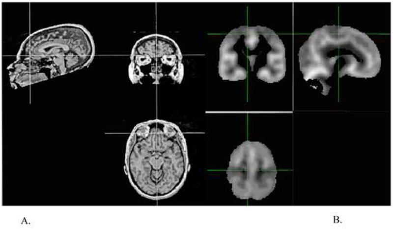
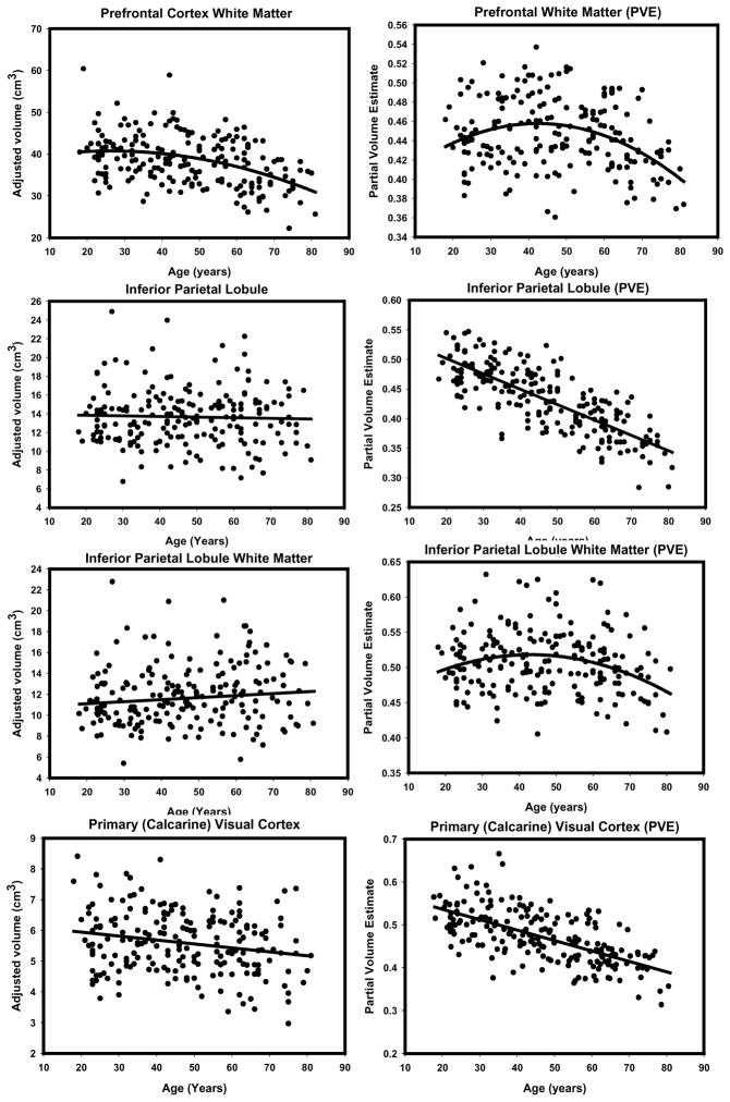
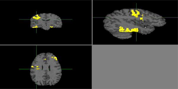
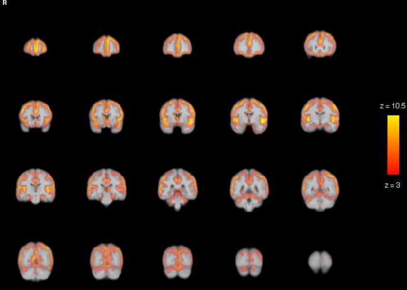
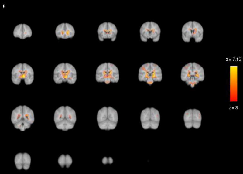
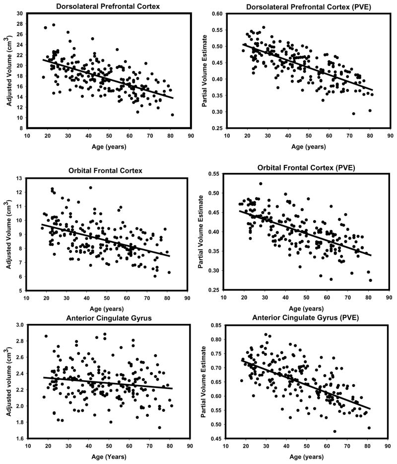
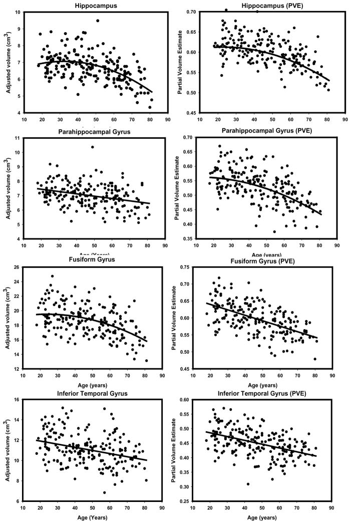
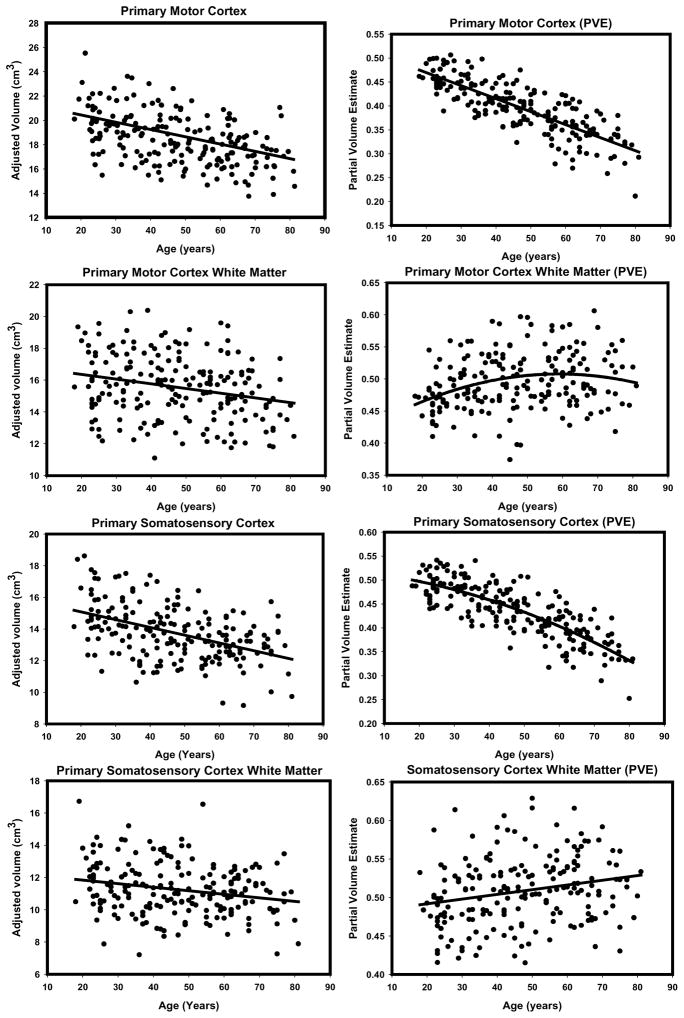
Regional manual volumetry is the gold standard of in vivo neuroanatomy, but is labor-intensive, can be imperfectly reliable, and allows for measuring limited number of regions. Voxel-based morphometry (VBM) has perfect repeatability and assesses local structure across the whole brain. However, its anatomic validity is unclear, and with its increasing popularity, a systematic comparison of VBM to manual volumetry is necessary. The few existing comparison studies are limited by small samples, qualitative comparisons, and limited selection and modest reliability of manual measures. Our goal was to overcome those limitations by quantitatively comparing optimized VBM findings with highly reliable multiple regional measures in a large sample (N=200) across a wide agespan (18-81). We report a complex pattern of similarities and differences. Peak values of VBM volume estimates (modulated density) produced stronger age differences and a different spatial distribution from manual measures. However, when we aggregated VBM-derived information across voxels contained in specific anatomically defined regions (masks), the patterns of age differences became more similar, although important discrepancies emerged. Notably, VBM revealed stronger age differences in the regions bordering CSF and white matter areas prone to leukoaraiosis, and VBM was more likely to report nonlinearities in age-volume relationships. In the white matter regions, manual measures showed stronger negative associations with age than the corresponding VBM-based masks. We conclude that VBM provides realistic estimates of age differences in the regional gray matter only when applied to anatomically defined regions, but overestimates effects when individual peaks are interpreted. It may be beneficial to use VBM as a first-pass strategy, followed by manual measurement of anatomically defined regions.
局部手动容积测量法是活体神经解剖学的金标准,但它劳动强度大,可靠性可能欠佳,且能测量的区域数量有限。基于体素的形态测量法(VBM)具有完美的可重复性,可评估全脑的局部结构。然而,其解剖学有效性尚不清楚,随着其日益普及,有必要对VBM与手动容积测量法进行系统比较。现有的少数比较研究受到样本量小、定性比较以及手动测量选择有限和可靠性一般的限制。我们的目标是通过在大样本(N = 200)、跨广泛年龄范围(18 - 81岁)的情况下,将优化后的VBM结果与高度可靠的多个区域测量结果进行定量比较,来克服这些限制。我们报告了相似性和差异的复杂模式。VBM体积估计值(调制密度)的峰值产生的年龄差异比手动测量更强,且空间分布不同。然而,当我们汇总特定解剖学定义区域(掩码)中包含的体素的VBM衍生信息时,年龄差异模式变得更加相似,尽管出现了重要差异。值得注意的是,VBM在与脑脊液相邻的区域以及易发生脑白质疏松的白质区域显示出更强的年龄差异,并且VBM更有可能报告年龄 - 体积关系中的非线性。在白质区域,手动测量显示出比相应的基于VBM的掩码与年龄更强的负相关。我们得出结论,VBM仅在应用于解剖学定义区域时才能提供区域灰质年龄差异的实际估计,但在解释单个峰值时会高估影响。将VBM用作初步策略,随后对手动测量解剖学定义区域可能是有益的。