Larouche Matt, Beffert Uwe, Herz Joachim, Hawkes Richard
Genes and Development Research Group and Hotchkiss Brain Institute, Department of Cell Biology and Anatomy, Faculty of Medicine, The University of Calgary, Calgary, Alberta, Canada.
PLoS One. 2008 Feb 27;3(2):e1653. doi: 10.1371/journal.pone.0001653.
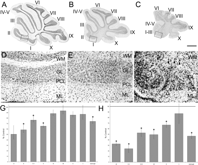
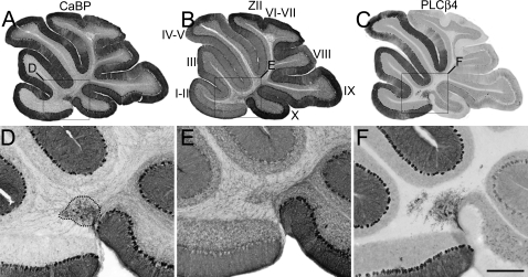
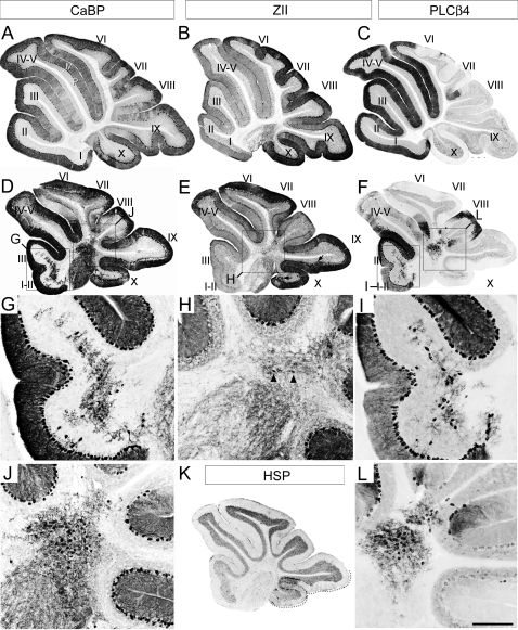
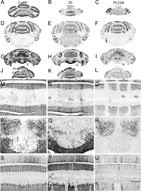
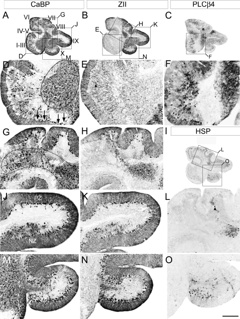
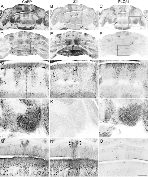
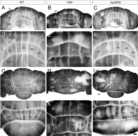
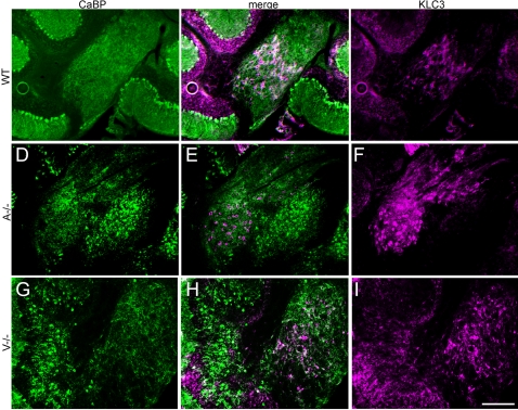
The adult cerebellar cortex is comprised of reproducible arrays of transverse zones and parasagittal stripes of Purkinje cells. Adult stripes are created through the perinatal rostrocaudal dispersion of embryonic Purkinje cell clusters, triggered by signaling through the Reelin pathway. Reelin is secreted by neurons in the external granular layer and deep cerebellar nuclei and binds to two high affinity extracellular receptors on Purkinje cells-the Very low density lipoprotein receptor (Vldlr) and apolipoprotein E receptor 2 (Apoer2). In mice null for either Reelin or double null for Vldlr and Apoer2, Purkinje cell clusters fail to disperse. Here we report that animals null for either Vldlr or Apoer2 individually, exhibit specific and parasagittally-restricted Purkinje cell ectopias. For example, in mice lacking Apoer2 function immunostaining reveals ectopic Purkinje cells that are largely restricted to the zebrin II-immunonegative population of the anterior vermis. In contrast, mice null for Vldlr have a much larger population of ectopic Purkinje cells that includes members from both the zebrin II-immunonegative and -immunopositive phenotypes. HSP25 immunoreactivity reveals that in Vldlr null animals a large portion of zebrin II-immunopositive ectopic cells are probably destined to become stripes in the central zone (lobules VI-VII). A small population of ectopic zebrin II-immunonegative Purkinje cells is also observed in animals heterozygous for both receptors (Apoer2(+/-): Vldlr(+/-)), but no ectopia is present in mice heterozygous for either receptor alone. These results indicate that Apoer2 and Vldlr coordinate the dispersal of distinct, but overlapping subsets of Purkinje cells in the developing cerebellum.
成年小脑皮质由浦肯野细胞的可重复排列的横向区域和矢状旁条纹组成。成年条纹是由胚胎浦肯野细胞簇在围产期的头尾向分散形成的,这一过程由Reelin信号通路触发。Reelin由外颗粒层和小脑深部核团中的神经元分泌,并与浦肯野细胞上的两种高亲和力细胞外受体——极低密度脂蛋白受体(Vldlr)和载脂蛋白E受体2(Apoer2)结合。在Reelin基因敲除小鼠或Vldlr和Apoer2双基因敲除小鼠中,浦肯野细胞簇无法分散。在这里,我们报告称,单独缺失Vldlr或Apoer2的动物表现出特定的、矢状旁受限的浦肯野细胞异位。例如,在缺乏Apoer2功能的小鼠中,免疫染色显示异位浦肯野细胞主要局限于前叶蚓部的zebrin II免疫阴性群体。相比之下,Vldlr基因敲除小鼠有更多的异位浦肯野细胞群体,包括zebrin II免疫阴性和免疫阳性表型的成员。HSP25免疫反应性显示,在Vldlr基因敲除动物中,大部分zebrin II免疫阳性异位细胞可能注定会在中央区(小叶VI-VII)形成条纹。在两种受体均为杂合子的动物(Apoer2(+/-): Vldlr(+/-))中也观察到一小部分异位的zebrin II免疫阴性浦肯野细胞,但单独一种受体为杂合子的小鼠中没有异位现象。这些结果表明,Apoer2和Vldlr在发育中的小脑中协调不同但重叠的浦肯野细胞亚群的分散。