Jennewein Marc, Lewis Matthew A, Zhao Dawen, Tsyganov Edward, Slavine Nikolai, He Jin, Watkins Linda, Kodibagkar Vikram D, O'Kelly Sean, Kulkarni Padmakar, Antich Peter P, Hermanne Alex, Rösch Frank, Mason Ralph P, Thorpe Philip E
Institute of Nuclear Chemistry, Johannes Gutenberg-University of Mainz, Mainz, Germany.
Clin Cancer Res. 2008 Mar 1;14(5):1377-85. doi: 10.1158/1078-0432.CCR-07-1516.
We recently reported that anionic phospholipids, principally phosphatidylserine, become exposed on the external surface of vascular endothelial cells in tumors, probably in response to oxidative stresses present in the tumor microenvironment. In the present study, we tested the hypothesis that a chimeric monoclonal antibody that binds phosphatidylserine could be labeled with radioactive arsenic isotopes and used for molecular imaging of solid tumors in rats.
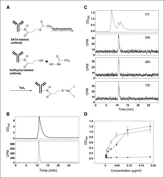
Bavituximab was labeled with (74)As (beta(+), T(1/2) 17.8 days) or (77)As (beta(-), T(1/2) 1.6 days) using a novel procedure. The radionuclides of arsenic were selected because their long half-lives are consistent with the long biological half lives of antibodies in vivo and because their chemistry permits stable attachment to antibodies. The radiolabeled antibodies were tested for the ability to image subcutaneous Dunning prostate R3227-AT1 tumors in rats.
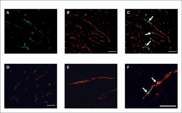
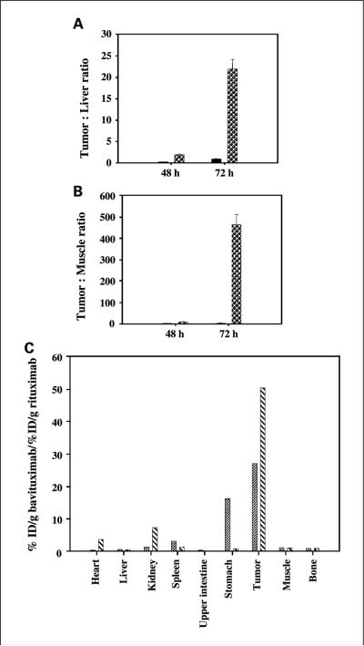
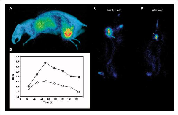
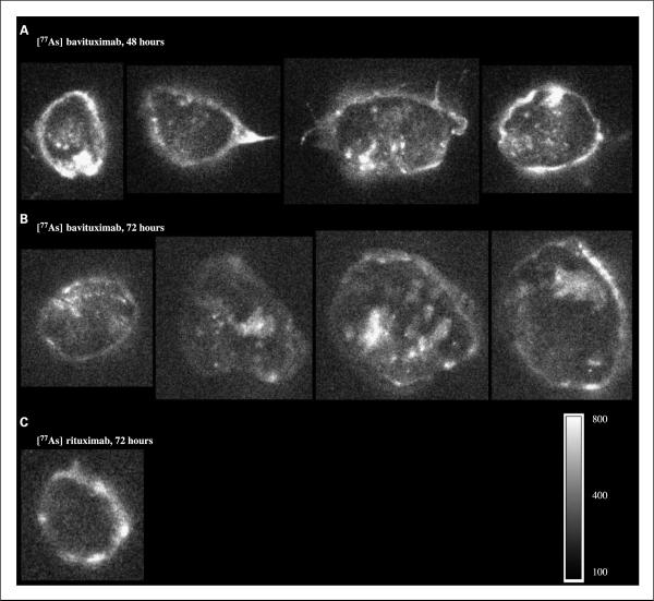
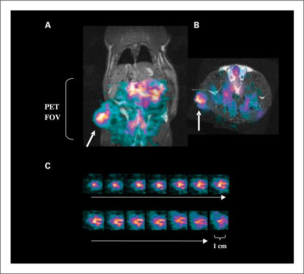
Clear images of the tumors were obtained using planar gamma-scintigraphy and positron emission tomography. Biodistribution studies confirmed the specific localization of bavituximab to the tumors. The tumor-to-liver ratio 72 h after injection was 22 for bavituximab compared with 1.5 for an isotype-matched control chimeric antibody of irrelevant specificity. Immunohistochemical studies showed that the bavituximab was labeling the tumor vascular endothelium.
These results show that radioarsenic-labeled bavituximab has potential as a new tool for imaging the vasculature of solid tumors.
我们最近报道,阴离子磷脂,主要是磷脂酰丝氨酸,在肿瘤血管内皮细胞的外表面暴露,这可能是对肿瘤微环境中存在的氧化应激的反应。在本研究中,我们测试了这样一个假设,即一种结合磷脂酰丝氨酸的嵌合单克隆抗体可以用放射性砷同位素标记,并用于大鼠实体瘤的分子成像。
使用一种新方法将巴维昔单抗用(74)As(β+,半衰期17.8天)或(77)As(β-,半衰期1.6天)标记。选择砷的放射性核素是因为它们的长半衰期与抗体在体内的长生物半衰期一致,并且因为它们的化学性质允许稳定地附着于抗体。对放射性标记的抗体进行成像大鼠皮下邓宁前列腺R3227-AT1肿瘤的能力测试。
使用平面γ闪烁扫描和正电子发射断层扫描获得了肿瘤的清晰图像。生物分布研究证实了巴维昔单抗在肿瘤中的特异性定位。注射后72小时,巴维昔单抗的肿瘤与肝脏比值为22,而具有无关特异性的同型匹配对照嵌合抗体的该比值为1.5。免疫组织化学研究表明,巴维昔单抗标记肿瘤血管内皮。
这些结果表明,放射性砷标记的巴维昔单抗有潜力作为一种成像实体瘤脉管系统的新工具。