Walker J, Jackson H, Brandon M R, Meeusen E
Centre for Animal Biotechnology, School of Veterinary Science, University of Melbourne, Parkville, Victoria, Australia.
Clin Exp Immunol. 1991 Oct;86(1):13-8. doi: 10.1111/j.1365-2249.1991.tb05766.x.
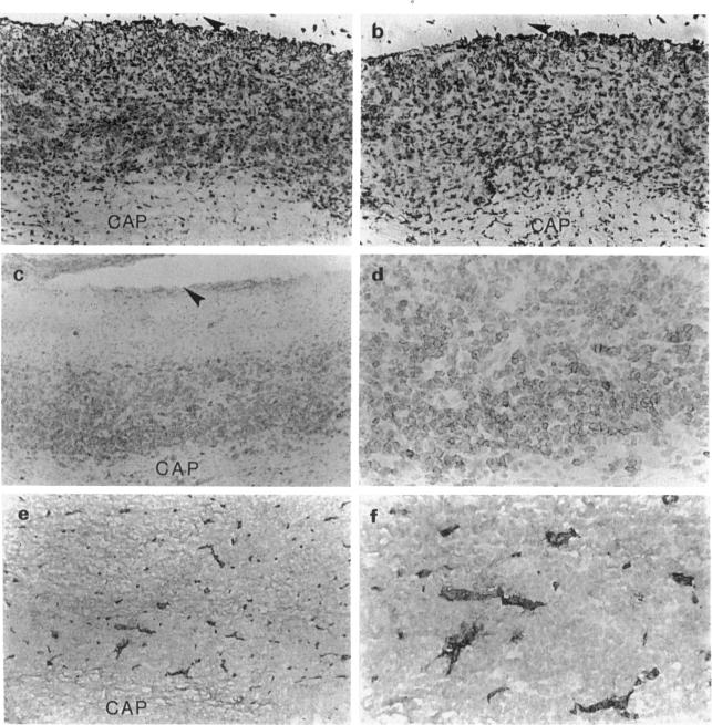
Pyogranulomas of ovine caseous lymphadenitis (CLA) are encapsulated lesions resulting from infections with Corynebacterium pseudotuberculosis, a bacterial pathogen able to grow within macrophages. Immunohistology of CLA lesions showed a band of lymphocytes lining the inside of the collagen capsule in intimate contact with necrotic tissue, the intracapsular lymphocytes being organized into three layers. The innermost layer, immediately adjacent to the central necrotic tissue consisted of a narrow band of MHC class II staining macrophages. Cells staining for CD4, CD8 and gamma delta T cell markers were unevenly distributed throughout the lymphoid layer, tending to be more numerous immediately external to the macrophage layer. The intracapsular lymphoid tissue contained a high proportion of CD8+ lymphocytes (CD4:CD8, 1.5:1) and of gamma delta lymphocytes (CD4:CD8:gamma delta, 1:0.7:0.8). External to the T cell-rich zone and adjacent to the surrounding collagen capsule was a dense band of cells, a proportion of which stained atypically for CD45R and were tentatively identified as B cells. CD8+ and gamma delta+ T cells showed similar distributions and their relative abundance, compared with CD4+ T cells, was a distinguishing feature of the CLA lesion. Staining for factor VIII-related antigen clearly showed endothelial venules throughout the intracapsular lymphoid tissue. The presence of endothelial venules and the organized architecture of the lymphoid tissue teleologically argues that lymphocytes are continually recruited into chronic CLA lesions and play an important role in the ongoing disease process.
绵羊干酪性淋巴结炎(CLA)的脓性肉芽肿是由伪结核棒状杆菌感染引起的包膜性病变,伪结核棒状杆菌是一种能够在巨噬细胞内生长的细菌病原体。CLA病变的免疫组织学显示,一层淋巴细胞排列在胶原包膜内部,与坏死组织紧密接触,包膜内的淋巴细胞分为三层。最内层紧邻中央坏死组织,由一条狭窄的MHC II类染色巨噬细胞带组成。CD4、CD8和γδ T细胞标志物染色的细胞在整个淋巴层中分布不均,在巨噬细胞层外侧数量往往更多。包膜内淋巴组织中CD8+淋巴细胞比例较高(CD4:CD8为1.5:1),γδ淋巴细胞比例也较高(CD4:CD8:γδ为1:0.7:0.8)。在富含T细胞的区域外侧且紧邻周围胶原包膜的是一条密集的细胞带,其中一部分细胞CD45R染色异常,初步鉴定为B细胞。CD8+和γδ+ T细胞分布相似,与CD4+ T细胞相比,它们的相对丰度是CLA病变的一个显著特征。VIII因子相关抗原染色清楚地显示了包膜内淋巴组织中的内皮微静脉。内皮微静脉的存在以及淋巴组织的有组织架构从目的论角度表明,淋巴细胞不断被招募到慢性CLA病变中,并在持续的疾病过程中发挥重要作用。