Cohen Alexander L, Fair Damien A, Dosenbach Nico U F, Miezin Francis M, Dierker Donna, Van Essen David C, Schlaggar Bradley L, Petersen Steven E
Department of Neurology, Washington University School of Medicine, St. Louis, MO 63110, USA.
Neuroimage. 2008 May 15;41(1):45-57. doi: 10.1016/j.neuroimage.2008.01.066. Epub 2008 Mar 25.
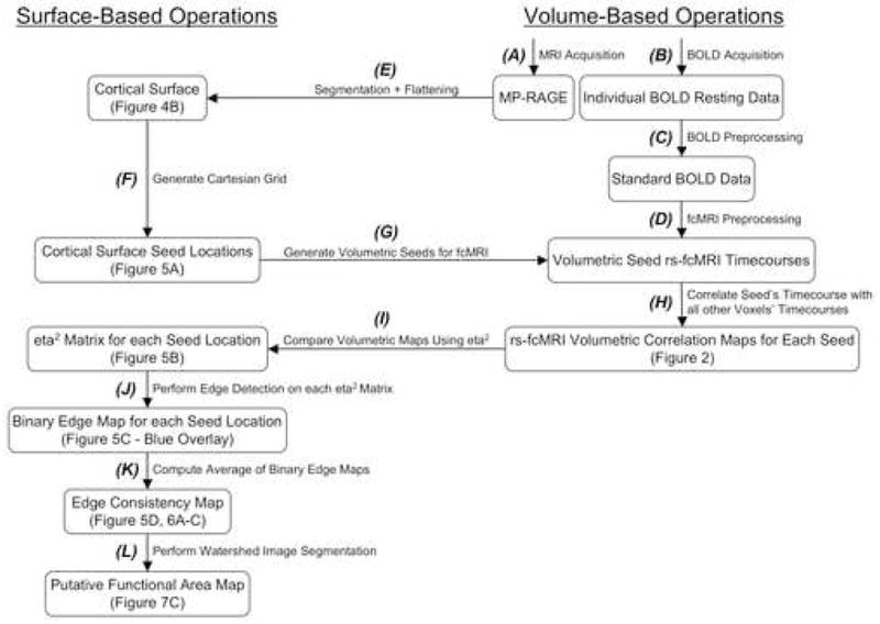
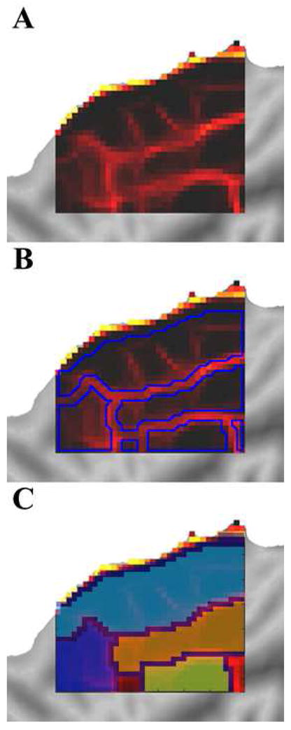
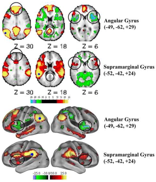
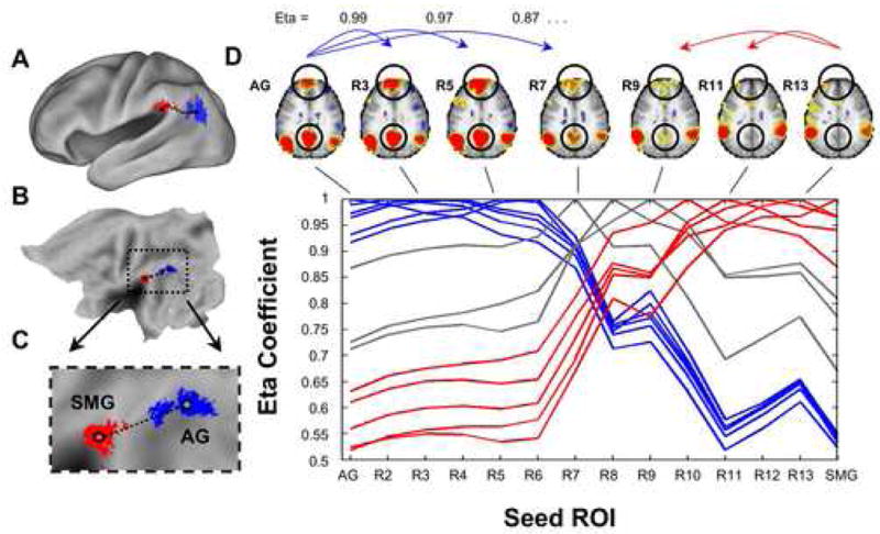
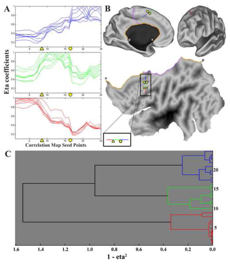
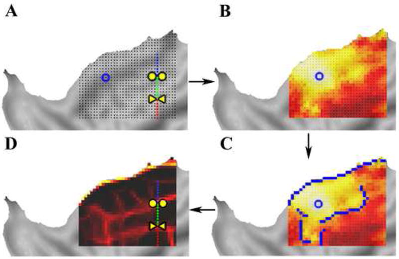
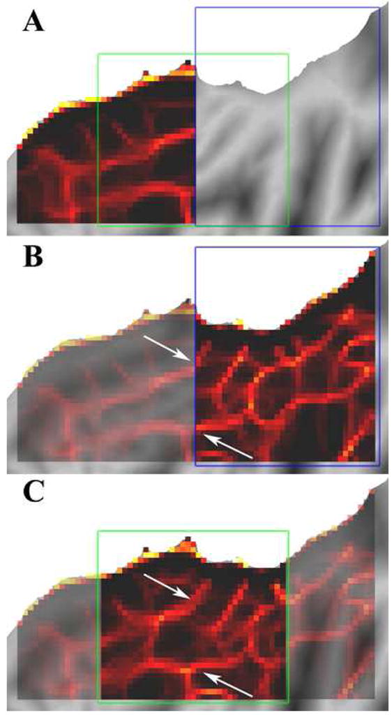
The cerebral cortex is anatomically organized at many physical scales starting at the level of single neurons and extending up to functional systems. Current functional magnetic resonance imaging (fMRI) studies often focus at the level of areas, networks, and systems. Except in restricted domains, (e.g., topographically-organized sensory regions), it is difficult to determine area boundaries in the human brain using fMRI. The ability to delineate functional areas non-invasively would enhance the quality of many experimental analyses allowing more accurate across-subject comparisons of independently identified functional areas. Correlations in spontaneous BOLD activity, often referred to as resting state functional connectivity (rs-fcMRI), are especially promising as a way to accurately localize differences in patterns of activity across large expanses of cortex. In the current report, we applied a novel set of image analysis tools to explore the utility of rs-fcMRI for defining wide-ranging functional area boundaries. We find that rs-fcMRI patterns show sharp transitions in correlation patterns and that these putative areal boundaries can be reliably detected in individual subjects as well as in group data. Additionally, combining surface-based analysis techniques with image processing algorithms allows automated mapping of putative areal boundaries across large expanses of cortex without the need for prior information about a region's function or topography. Our approach reliably produces maps of bounded regions appropriate in size and number for putative functional areas. These findings will hopefully stimulate further methodological refinements and validations.
大脑皮层在解剖学上是按照从单个神经元水平开始直至功能系统的多个物理尺度进行组织的。当前的功能磁共振成像(fMRI)研究通常聚焦于区域、网络和系统层面。除了在受限的领域(例如,具有拓扑组织的感觉区域),使用fMRI很难确定人类大脑中的区域边界。非侵入性地描绘功能区域的能力将提高许多实验分析的质量,从而允许对独立识别的功能区域进行更准确的跨个体比较。自发血氧水平依赖(BOLD)活动中的相关性,通常被称为静息态功能连接(rs-fcMRI),作为一种准确定位大片皮层活动模式差异的方法尤其具有前景。在本报告中,我们应用了一组新颖的图像分析工具来探索rs-fcMRI在定义广泛的功能区域边界方面的效用。我们发现rs-fcMRI模式在相关模式中显示出急剧转变,并且这些假定的区域边界能够在个体受试者以及群体数据中被可靠地检测到。此外,将基于表面的分析技术与图像处理算法相结合,可以在无需关于区域功能或地形的先验信息的情况下,自动绘制大片皮层上假定的区域边界。我们的方法能够可靠地生成大小和数量适合假定功能区域的有界区域图谱。这些发现有望激发进一步的方法学改进和验证。