Sir Peter Mansfield Magnetic Resonance Centre, School of Physics and Astronomy, University of Nottingham, University Park, Nottingham, UK.
Neuroimage. 2011 Jun 1;56(3):1082-104. doi: 10.1016/j.neuroimage.2011.02.054. Epub 2011 Feb 23.
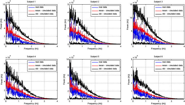
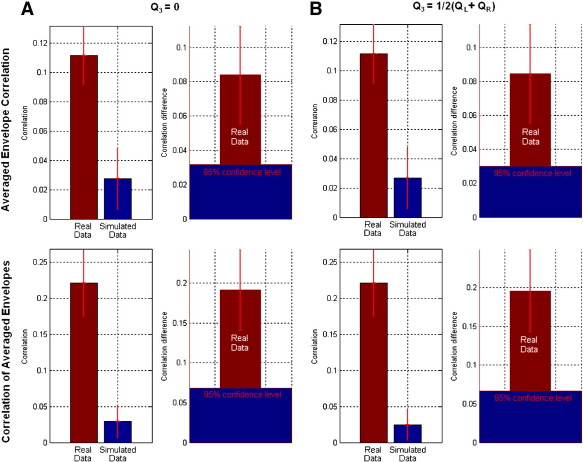
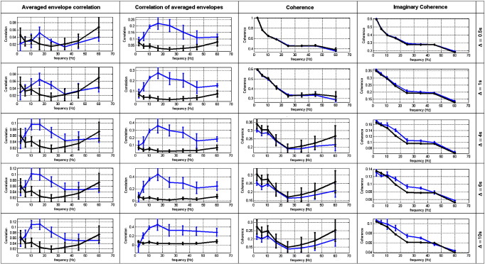
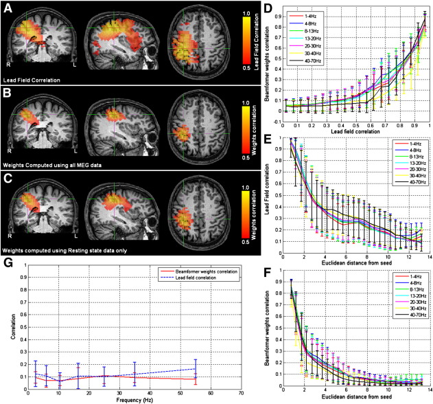
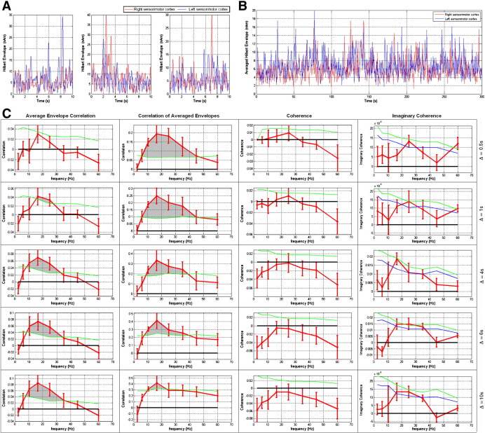
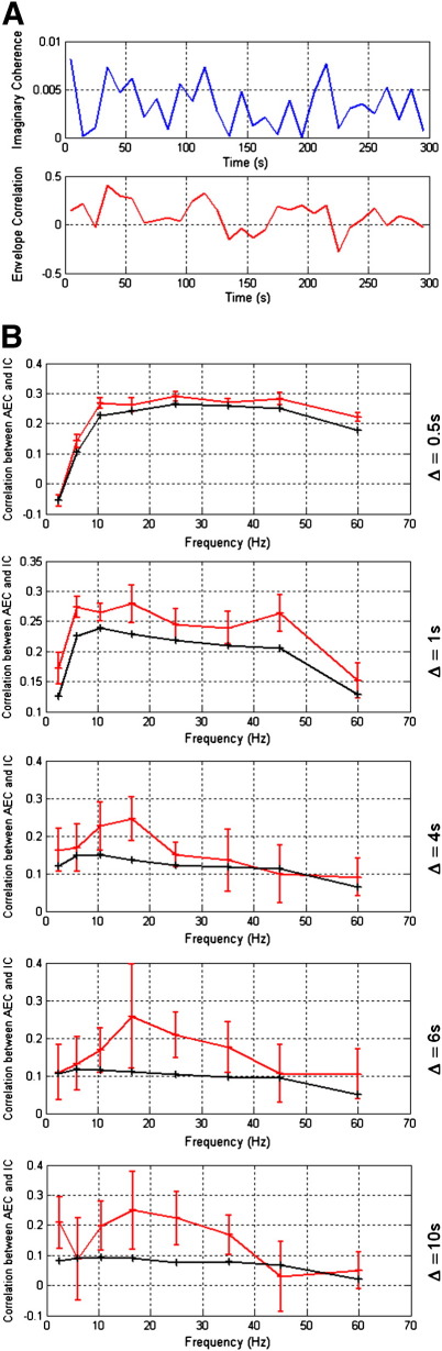
Functional connectivity (FC) between brain regions is thought to be central to the way in which the brain processes information. Abnormal connectivity is thought to be implicated in a number of diseases. The ability to study FC is therefore a key goal for neuroimaging. Functional connectivity (fc) MRI has become a popular tool to make connectivity measurements but the technique is limited by its indirect nature. A multimodal approach is therefore an attractive means to investigate the electrodynamic mechanisms underlying hemodynamic connectivity. In this paper, we investigate resting state FC using fcMRI and magnetoencephalography (MEG). In fcMRI, we exploit the advantages afforded by ultra high magnetic field. In MEG we apply envelope correlation and coherence techniques to source space projected MEG signals. We show that beamforming provides an excellent means to measure FC in source space using MEG data. However, care must be taken when interpreting these measurements since cross talk between voxels in source space can potentially lead to spurious connectivity and this must be taken into account in all studies of this type. We show good spatial agreement between FC measured independently using MEG and fcMRI; FC between sensorimotor cortices was observed using both modalities, with the best spatial agreement when MEG data are filtered into the β band. This finding helps to reduce the potential confounds associated with each modality alone: while it helps reduce the uncertainties in spatial patterns generated by MEG (brought about by the ill posed inverse problem), addition of electrodynamic metric confirms the neural basis of fcMRI measurements. Finally, we show that multiple MEG based FC metrics allow the potential to move beyond what is possible using fcMRI, and investigate the nature of electrodynamic connectivity. Our results extend those from previous studies and add weight to the argument that neural oscillations are intimately related to functional connectivity and the BOLD response.
功能连接(FC)被认为是大脑处理信息的核心方式。异常的连接被认为与许多疾病有关。因此,研究 FC 的能力是神经影像学的一个关键目标。功能连接(fc)MRI 已成为一种流行的工具来进行连接测量,但该技术受到其间接性质的限制。因此,多模态方法是研究血流动力学连接背后的电磁动力学机制的一种有吸引力的手段。在本文中,我们使用 fcMRI 和脑磁图(MEG)研究静息状态 FC。在 fcMRI 中,我们利用超高磁场的优势。在 MEG 中,我们应用包络相关和相干技术来处理源空间投影的 MEG 信号。我们表明,波束形成提供了一种极好的方法,可以使用 MEG 数据在源空间中测量 FC。然而,在解释这些测量值时必须小心,因为源空间中的体素之间的串扰可能会导致虚假的连接,在这种类型的所有研究中都必须考虑到这一点。我们表明,使用 MEG 和 fcMRI 独立测量的 FC 之间具有良好的空间一致性;两种模态都观察到感觉运动皮质之间的 FC,当 MEG 数据被过滤到β频带时,空间一致性最佳。这一发现有助于减少每种模态单独存在的潜在混淆:虽然它有助于减少 MEG 产生的空间模式的不确定性(由不适定的逆问题引起),但添加电磁度量法确认了 fcMRI 测量的神经基础。最后,我们表明,多个基于 MEG 的 FC 指标有可能超越使用 fcMRI 可能实现的功能,并研究电磁连接的性质。我们的结果扩展了以前的研究结果,并为神经振荡与功能连接和 BOLD 反应密切相关的论点增加了份量。