Laughlin Scott T, Baskin Jeremy M, Amacher Sharon L, Bertozzi Carolyn R
Department of Chemistry, University of California, Berkeley, CA 94720, USA.
Science. 2008 May 2;320(5876):664-7. doi: 10.1126/science.1155106.
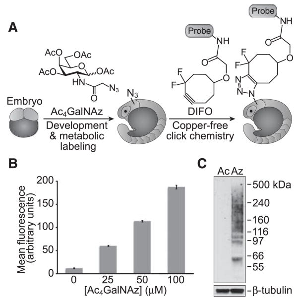
Glycans are attractive targets for molecular imaging but have been inaccessible because of their incompatibility with genetically encoded reporters. We demonstrated the noninvasive imaging of glycans in live developing zebrafish, using a chemical reporter strategy. Zebrafish embryos were treated with an unnatural sugar to metabolically label their cell-surface glycans with azides. Subsequently, the embryos were reacted with fluorophore conjugates by means of copper-free click chemistry, enabling the visualization of glycans in vivo at subcellular resolution during development. At 60 hours after fertilization, we observed an increase in de novo glycan biosynthesis in the jaw region, pectoral fins, and olfactory organs. Using a multicolor detection strategy, we performed a spatiotemporal analysis of glycan expression and trafficking and identified patterns that would be undetectable with conventional molecular imaging approaches.
聚糖是分子成像的诱人靶点,但由于它们与基因编码报告分子不兼容,一直难以实现成像。我们采用化学报告策略,在活体发育斑马鱼中实现了聚糖的无创成像。用一种非天然糖处理斑马鱼胚胎,以代谢方式用叠氮化物标记其细胞表面聚糖。随后,通过无铜点击化学使胚胎与荧光团共轭物反应,从而在发育过程中以亚细胞分辨率在体内实现聚糖的可视化。受精后60小时,我们观察到颌部区域、胸鳍和嗅觉器官中从头聚糖生物合成增加。利用多色检测策略,我们对聚糖表达和运输进行了时空分析,并确定了传统分子成像方法无法检测到的模式。