Jiang Hao, Zheng Tianqing, Lopez-Aguilar Aime, Feng Lei, Kopp Felix, Marlow Florence L, Wu Peng
Department of Biochemistry, ‡Chemical Biology Core Facility and §Developmental and Molecular Biology, Albert Einstein College of Medicine, Yeshiva University , 1300 Morris Park Avenue, Bronx, New York 10461, United States.
Bioconjug Chem. 2014 Apr 16;25(4):698-706. doi: 10.1021/bc400502d. Epub 2014 Mar 14.
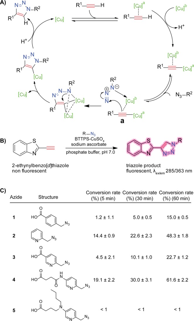
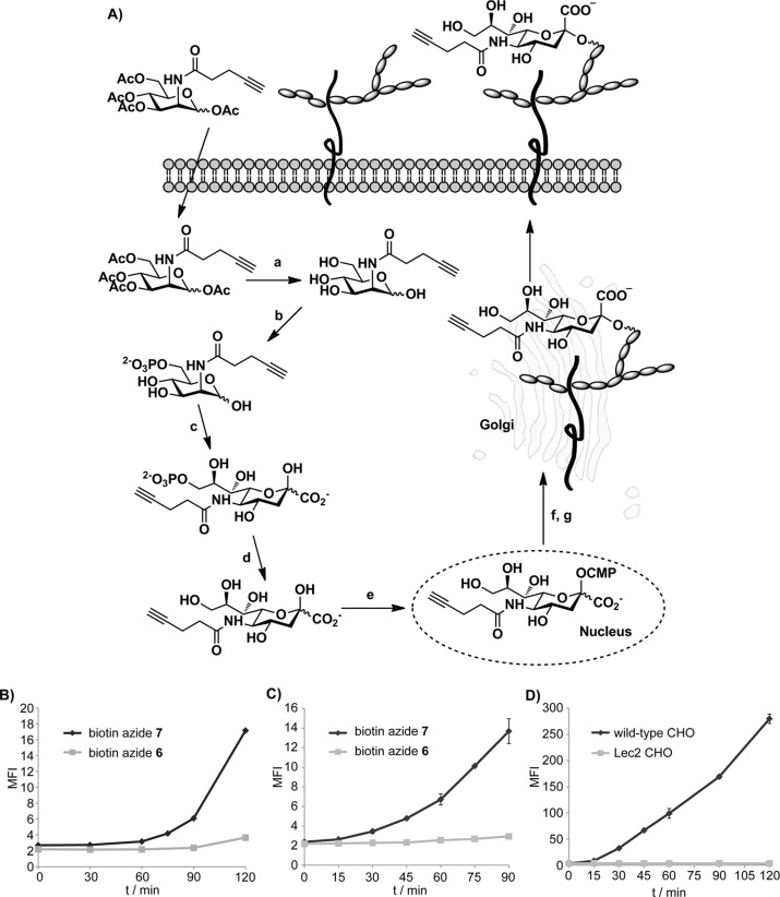
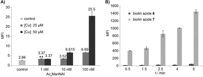
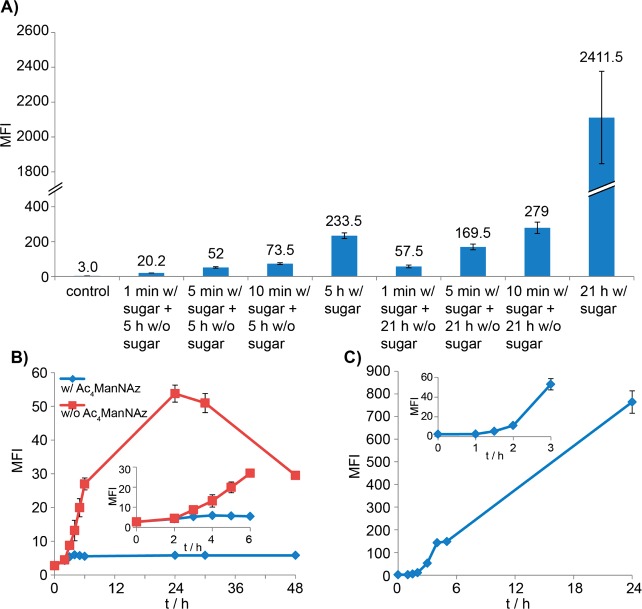
To monitor the kinetics of biological processes that take place within the minute time scale, simple and fast analytical methods are required. In this article, we present our discovery of an azide with an internal Cu(I)-chelating motif that enabled the development of the fastest protocol for Cu(I)-catalyzed azide-alkyne cycloaddition (CuAAC) to date, and its application toward following the dynamic process of glycan biosynthesis. We discovered that an electron-donating picolyl azide boosted the efficiency of the ligand-accelerated CuAAC 20-38-fold in living systems with no apparent toxicity. With a combination of this azide and BTTPS, a tris(triazolylmethyl)amine-based ligand for Cu(I), we were able to detect newly synthesized cell-surface glycans by flow cytometry using as low as 1 nM of a metabolic precursor. This supersensitive chemistry enabled us to monitor the dynamic glycan biosynthesis in mammalian cells and in early zebrafish embryogenesis. In live mammalian cells, we discovered that it takes approximately 30-45 min for a monosaccharide building block to be metabolized and incorporated into cell-surface glycoconjugates. In zebrafish embryos, the labeled glycans could be detected as early as the two-cell stage. To our knowledge, this was the first time that newly synthesized glycans were detected at the cleavage period (0.75-2 hpf) in an animal model using bioorthogonal chemistry.
为了监测在分钟时间尺度内发生的生物过程的动力学,需要简单快速的分析方法。在本文中,我们展示了我们发现的一种带有内部Cu(I)螯合基序的叠氮化物,它使得开发出了迄今为止用于Cu(I)催化的叠氮化物-炔烃环加成反应(CuAAC)的最快方案,以及其在追踪聚糖生物合成动态过程中的应用。我们发现,供电子的吡啶甲基叠氮化物在无明显毒性的活系统中将配体加速的CuAAC效率提高了20至38倍。将这种叠氮化物与用于Cu(I)的基于三(三唑基甲基)胺的配体BTTPS相结合,我们能够通过流式细胞术使用低至1 nM的代谢前体来检测新合成的细胞表面聚糖。这种超灵敏化学方法使我们能够监测哺乳动物细胞和早期斑马鱼胚胎发育中的动态聚糖生物合成。在活的哺乳动物细胞中,我们发现一个单糖构建块代谢并掺入细胞表面糖缀合物大约需要30 - 45分钟。在斑马鱼胚胎中,最早在两细胞阶段就能检测到标记的聚糖。据我们所知,这是首次使用生物正交化学在动物模型的卵裂期(0.75 - 2 hpf)检测到新合成的聚糖。