Uffen M P, Krijnen M R, Hoogendoorn R J, Strijkers G J, Everts V, Wuisman P I, Smit T H
Department of Orthopaedic Surgery, VU University Medical Center, Amsterdam, The Netherlands.
Eur Spine J. 2008 Aug;17(8):1006-11. doi: 10.1007/s00586-008-0689-7. Epub 2008 May 30.
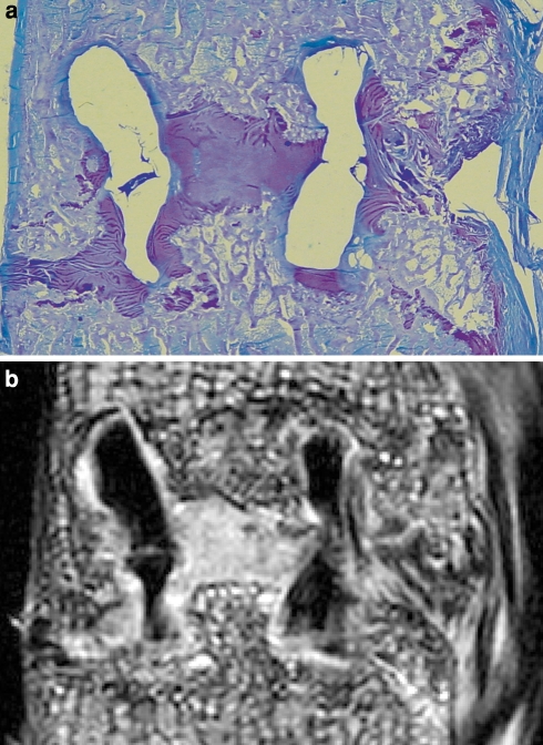
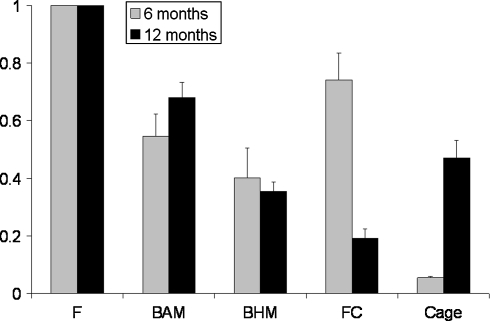
Nonunion is a major complication of spinal interbody fusion. Currently X-ray and computed tomography (CT) are used for evaluating the spinal fusion process. However, both imaging modalities have limitations in judgment of the early stages of this fusion process, as they only visualize mineralized bone. Magnetic resonance imaging (MRI) could be of great value as it is able to discriminate between different types of tissue. A feasibility study was performed in nine animals from a goat spinal fusion study, to evaluate the detection capacity of different tissues with micro-MRI. In this study bioresorbable polylactic acid cages were used. Six- and 12-months follow-up specimens were scanned in a 6.3 T micro-MRI scanner. After scanning, the specimens were processed for histology. Different types of tissue as well as the degradable cage material were identified in the fusion zone and designated as regions of interest (ROIs). Subsequently, the location of these ROIs was determined on the corresponding micro-MRI image, and average signal intensities of every individual ROI were measured. An excellent match was seen between the histological sections and micro-MRI images. The micro-MRI images showed quantifiable differences in signal intensity between bone with adipose marrow, bone with hematopoietic marrow, fibrocartilage, fibrous tissue, and degradable implant material. In time the signal intensity of bone with adipose marrow, bone with hematopoietic red marrow, and of fibrous tissue remained relatively constant. On the other hand, the signal intensity of the degradable implant material and the fibrocartilage changed significantly in time, indicating change of structure and composition. In conclusion, in our model using bioresorbable cages the MRI provides us with detailed information about the early fusion process and may therefore, allow early diagnosis of non-union.
骨不连是脊柱椎间融合术的一种主要并发症。目前,X射线和计算机断层扫描(CT)用于评估脊柱融合过程。然而,这两种成像方式在判断该融合过程的早期阶段时都存在局限性,因为它们只能显示矿化骨。磁共振成像(MRI)可能具有很大价值,因为它能够区分不同类型的组织。在一项山羊脊柱融合研究的九只动物身上进行了一项可行性研究,以评估显微MRI对不同组织的检测能力。在本研究中使用了可生物吸收的聚乳酸椎间融合器。对6个月和12个月随访的标本在6.3T显微MRI扫描仪中进行扫描。扫描后,对标本进行组织学处理。在融合区识别出不同类型的组织以及可降解的融合器材料,并将其指定为感兴趣区域(ROI)。随后,在相应的显微MRI图像上确定这些ROI的位置,并测量每个单独ROI的平均信号强度。组织学切片和显微MRI图像之间显示出极佳的匹配。显微MRI图像显示,含有脂肪骨髓的骨、含有造血骨髓的骨、纤维软骨、纤维组织和可降解植入材料之间在信号强度上存在可量化的差异。随着时间的推移,含有脂肪骨髓的骨、含有造血红骨髓的骨以及纤维组织的信号强度保持相对恒定。另一方面,可降解植入材料和纤维软骨的信号强度随时间发生了显著变化,表明结构和成分发生了改变。总之,在我们使用可生物吸收融合器的模型中,MRI为我们提供了有关早期融合过程的详细信息,因此可能有助于早期诊断骨不连。