Downs J Crawford, Roberts Michael D, Burgoyne Claude F
Devers Eye Institute, Legacy Health System, Portland, Oregon 97232, USA.
Optom Vis Sci. 2008 Jun;85(6):425-35. doi: 10.1097/OPX.0b013e31817841cb.
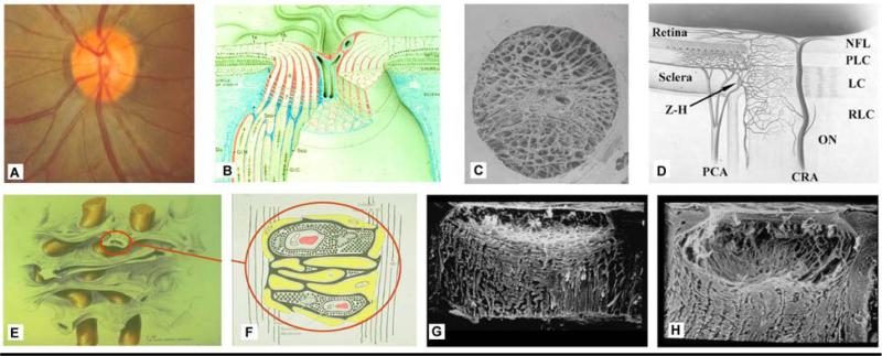
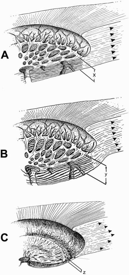
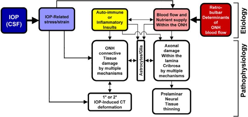
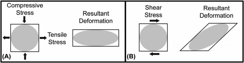
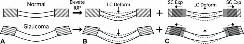
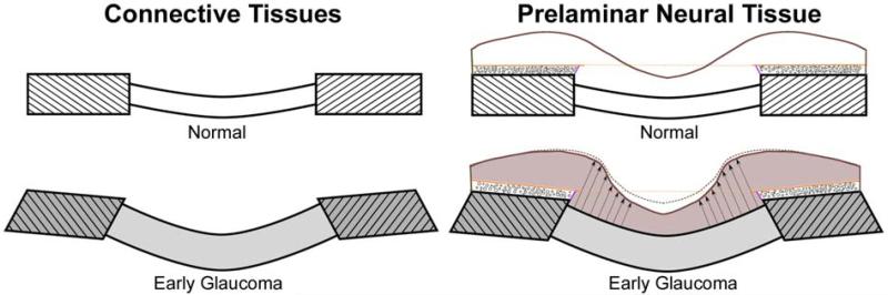
The optic nerve head (ONH) is of particular interest from a biomechanical perspective because it is a weak spot within an otherwise strong corneo-scleral envelope. The lamina cribrosa provides structural and functional support to the retinal ganglion cell axons as they pass from the relatively high-pressure environment in the eye to a low-pressure region in the retrobulbar cerebrospinal space. To protect the retinal ganglion cell axons within this unique environment, the lamina cribrosa in higher primates has developed into a complex structure composed of a three-dimensional network of flexible beams of connective tissue. The ONH is nourished by the short posterior ciliary arteries, which penetrate the immediate peripapillary sclera to feed capillaries contained within the laminar beams. This intrascleral and intralaminar vasculature is unique in that it is encased in load-bearing connective tissue, either within the scleral wall adjacent to the lamina cribrosa, or within the laminar beams themselves. Glaucoma is a multifactorial disease, and we believe that biomechanics not only determines the mechanical environment in the ONH, but also mediates IOP-related reductions in blood flow and cellular responses through various pathways. Our current understanding of the mechanical environment of the ONH is described, with particular emphasis on the influence of biomechanics in glaucoma.
从生物力学角度来看,视神经乳头(ONH)特别引人关注,因为它是原本坚固的角膜巩膜包膜中的一个薄弱点。筛板为视网膜神经节细胞轴突提供结构和功能支持,这些轴突从眼内相对高压的环境通向球后视神经脑脊液空间中的低压区域。为了在这种独特环境中保护视网膜神经节细胞轴突,高等灵长类动物的筛板已发展成为一种复杂结构,由结缔组织的柔性梁三维网络组成。视神经乳头由睫状后短动脉供血,这些动脉穿透紧邻视乳头周围的巩膜,为包含在板层梁内的毛细血管供血。这种巩膜内和板层内的脉管系统很独特,因为它被包裹在承重结缔组织中,要么在紧邻筛板的巩膜壁内,要么在板层梁本身内。青光眼是一种多因素疾病,我们认为生物力学不仅决定了视神经乳头内的机械环境,还通过各种途径介导与眼压相关的血流减少和细胞反应。本文描述了我们目前对视神经乳头机械环境的理解,特别强调了生物力学在青光眼中的影响。