Chang Nam Kyu, Jeong Yong Yeon, Park Jong Seong, Jeong Han Seong, Jang Sujeong, Jang Myung Joo, Lee Jae Hyuk, Shin Sang Soo, Yoon Woong, Chung Tae Woong, Kang Heoung Keun
Department of Radiology, Chonnam National University Medical School, Gwang-ju, Korea.
Korean J Radiol. 2008 May-Jun;9(3):196-204. doi: 10.3348/kjr.2008.9.3.196.
To access the feasibility of clinically available 3T MRI to detect the migration of labeled neural stem cells (NSCs) in intracerebral hemorrhage (ICH) in a rat model.
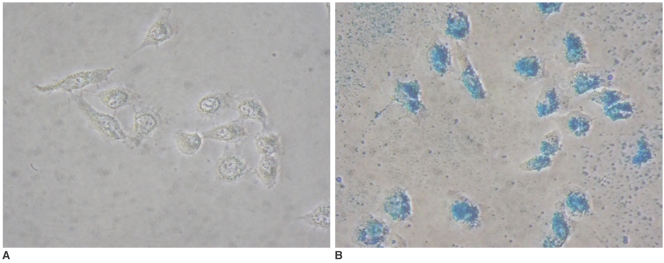
The ethics committee of our institution approved this study. ICH was induced by the injection of collagenase type IV into the right striatum of ten Sprague-Dawley rats. Human NSCs conjugated with Feridex (super-paramagnetic iron oxide: SPIO) were transplanted into the left striatum one week after ICH induction. MRI was performed on a 3T scanner during the first, second, third, fourth, and sixth weeks post-transplantation. MRI was obtained using coronal T2- and T2*-weighted sequences. Two rats were sacrificed every week after in vivo MRI in order to analyze the histological findings.
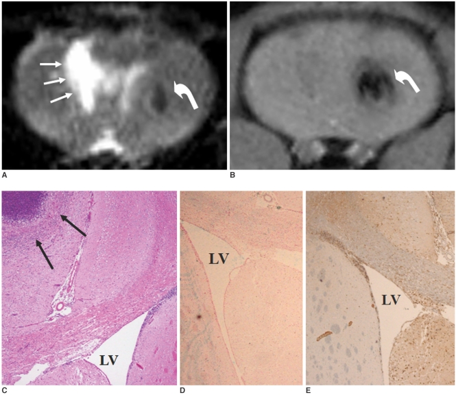
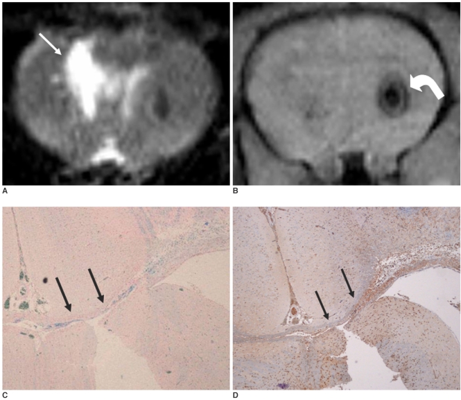
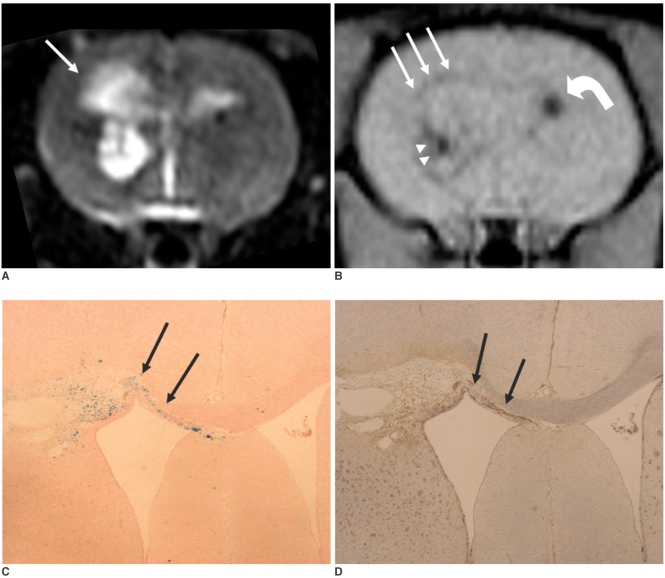
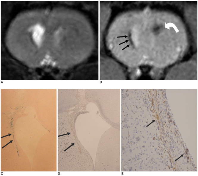
ICH in the right striatum was detected by MRI one and two weeks after transplantation without migration of the NSCs. There was no migration of the NSCs as seen on the histological findings one week after transplantation. The histological findings two weeks after transplantation showed a small number of NSCs along the corpus callosum. On MRI three weeks after transplantation, there was a hypointense line along the corpus callosum and decreased signal intensity in the right periventricular region. Histological findings three weeks after transplantation confirmed the presence of the hypointense line representing SPIO-labeled NSCs. MRI four and six weeks after transplantation showed a hypointense spot in the right periventricular region. The histological findings four and six weeks after transplantation showed the presence of prominent NSCs in the right periventricular region.
3T MRI can detect the migration of NSCs in rats with ICH along the corpus callosum. Therefore, 3T MRI could be feasible for detecting the migration of NSCs in the clinical setting of stem cell therapy.
探讨临床可用的3T磁共振成像(MRI)检测大鼠脑出血(ICH)模型中标记神经干细胞(NSCs)迁移的可行性。
本机构伦理委员会批准了本研究。通过向10只Sprague-Dawley大鼠的右侧纹状体注射IV型胶原酶诱导脑出血。在脑出血诱导1周后,将与菲立磁(超顺磁性氧化铁:SPIO)结合的人神经干细胞移植到左侧纹状体。在移植后的第1、2、3、4和6周,使用3T扫描仪进行MRI检查。采用冠状位T2加权和T2*加权序列获取MRI图像。在活体MRI检查后,每周处死2只大鼠以分析组织学结果。
移植后1周和2周,MRI检测到右侧纹状体有脑出血,未见神经干细胞迁移。移植后1周的组织学检查结果未见神经干细胞迁移。移植后2周的组织学检查结果显示胼胝体周围有少量神经干细胞。移植后3周的MRI检查显示胼胝体有一条低信号线,右侧脑室周围区域信号强度降低。移植后3周的组织学检查结果证实存在代表SPIO标记神经干细胞的低信号线。移植后4周和6周的MRI检查显示右侧脑室周围区域有一个低信号点。移植后4周和6周的组织学检查结果显示右侧脑室周围区域有大量神经干细胞。
3T MRI可检测ICH大鼠中神经干细胞沿胼胝体的迁移。因此,3T MRI在干细胞治疗的临床环境中检测神经干细胞的迁移可能是可行的。