Sibov Tatiana T, Pavon Lorena F, Miyaki Liza A, Mamani Javier B, Nucci Leopoldo P, Alvarim Larissa T, Silveira Paulo H, Marti Luciana C, Gamarra Lf
Hospital Israelita Albert Einstein, São Paulo, Brazil ; Departamento de Neurologia e Neurociências, Universidade Federal de São Paulo, São Paulo, Brazil.
Hospital Israelita Albert Einstein, São Paulo, Brazil.
Int J Nanomedicine. 2014 Jan 8;9:337-50. doi: 10.2147/IJN.S53299. eCollection 2014.
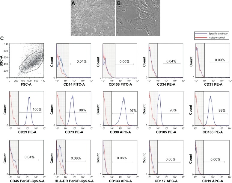
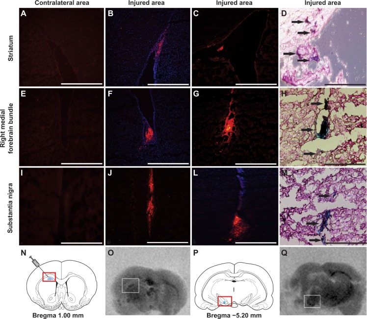
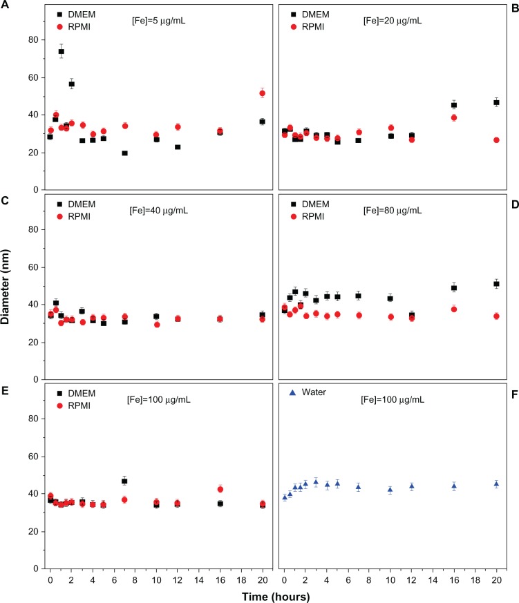
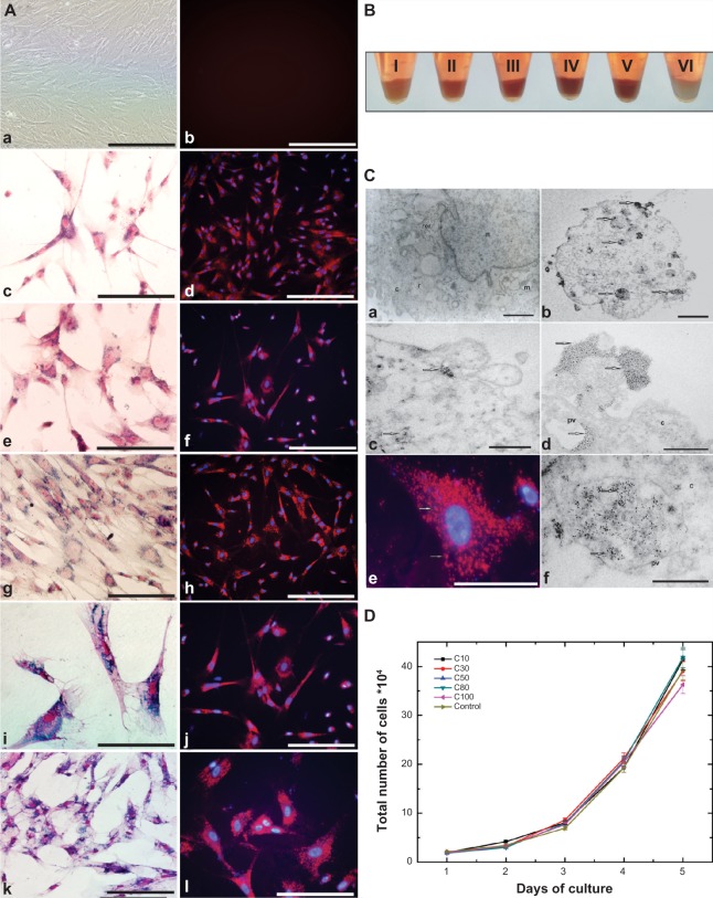
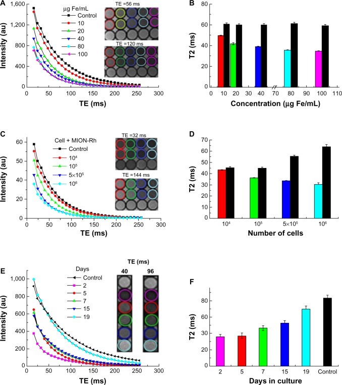
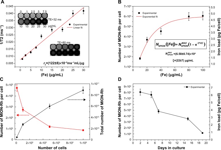
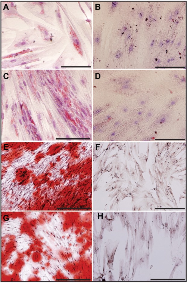
Here we describe multimodal iron oxide nanoparticles conjugated to Rhodamine-B (MION-Rh), their stability in culture medium, and subsequent validation of an in vitro protocol to label mesenchymal stem cells from umbilical cord blood (UC-MSC) with MION-Rh. These cells showed robust labeling in vitro without impairment of their functional properties, the viability of which were evaluated by proliferation kinetic and ultrastructural analyzes. Thus, labeled cells were infused into striatum of adult male rats of animal model that mimic late onset of Parkinson's disease and, after 15 days, it was observed that cells migrated along the medial forebrain bundle to the substantia nigra as hypointense spots in T2 magnetic resonance imaging. These data were supported by short-term magnetic resonance imaging. Studies were performed in vivo, which showed that about 5 × 10(5) cells could be efficiently detected in the short term following infusion. Our results indicate that these labeled cells can be efficiently tracked in a neurodegenerative disease model.
在此,我们描述了与罗丹明 - B 偶联的多模态氧化铁纳米颗粒(MION - Rh)、它们在培养基中的稳定性,以及随后用于用 MION - Rh 标记脐带血间充质干细胞(UC - MSC)的体外方案的验证。这些细胞在体外显示出强烈的标记,且其功能特性未受损害,通过增殖动力学和超微结构分析评估了它们的活力。因此,将标记的细胞注入模拟帕金森病晚期发病的成年雄性大鼠的纹状体中,15 天后,在 T2 磁共振成像中观察到细胞沿着内侧前脑束迁移至黑质,呈现为低信号点。这些数据得到了短期磁共振成像的支持。在体内进行的研究表明,输注后短期内可有效检测到约 5×10⁵个细胞。我们的结果表明,这些标记的细胞能够在神经退行性疾病模型中被有效追踪。