Rousset Olivier G, Collins D Louis, Rahmim Arman, Wong Dean F
Section of High Resolution Brain PET Imaging, Division of Nuclear Medicine, Russell H. Morgan Department of Radiology, Johns Hopkins University, Baltimore, Maryland 21287, USA.
J Nucl Med. 2008 Jul;49(7):1097-106. doi: 10.2967/jnumed.107.048330. Epub 2008 Jun 13.
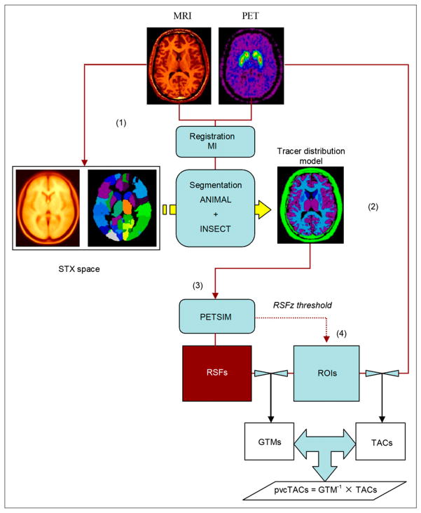
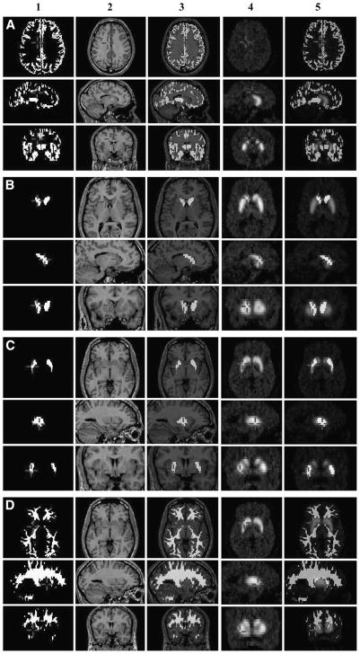
The considerable effort and potential lack of reproducibility of human-driven PET quantification and partial volume correction (PVC) can be alleviated by use of atlas-based automatic analysis. The present study examined the application of a new algorithm designed to automatically define 3-dimensional regions of interest (ROIs) and their effect on dopamine receptor quantification in the normal human brain striatum, both without and with PVC.
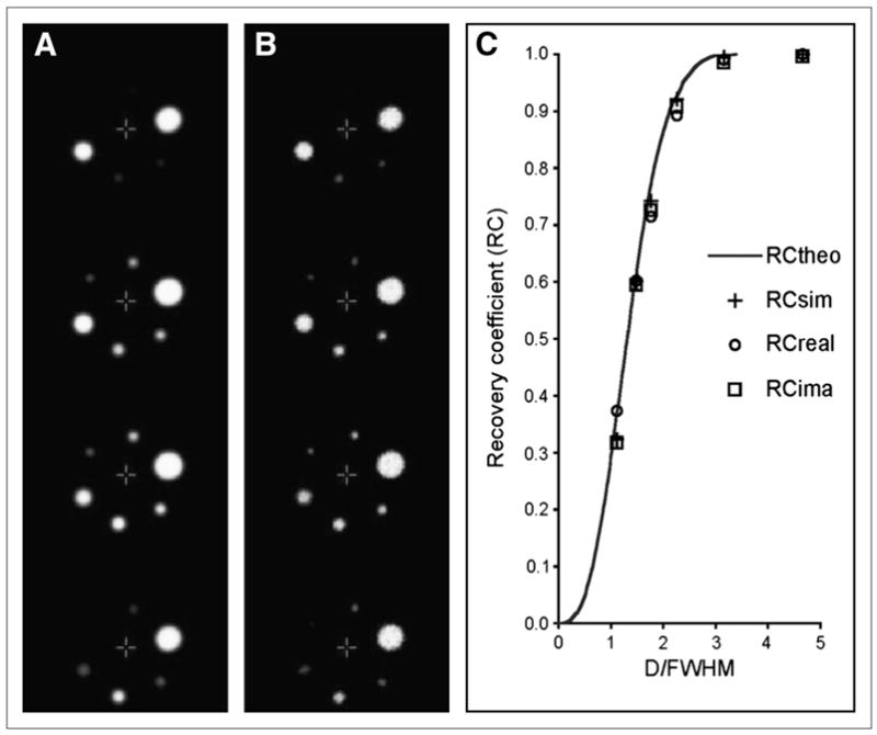
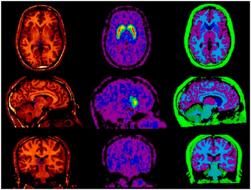
A total of 90 healthy volunteers (age range, 18-46 y) received a single injection of (11)C-raclopride, and automatic segmentation of concomitant structural MR images was performed using a maximum-probability atlas in combination with a trained neural network. For each identified tissue segment considered homogeneous for the tracer (or volumes of interest [VOIs]), an a priori criterion based on minimum axial recovery coefficient (RC(zmin) = 50%, 75%, and 90%) was used to constrain the extent of each ROI.
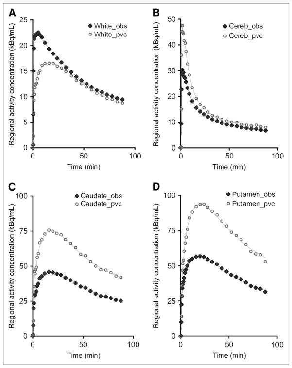
With ROIs essentially overlapping the entire VOI volume (obtained with RC(zmin) = 50%), the binding potential (BP(ND)) of (11)C-raclopride was found to be around 2.2 for caudate and 2.9 for putamen, an underestimation by 35% and 28%, respectively, according to PVC values. At increased RC(zmin), BP(ND) estimates of (11)C-raclopride were increased by 12% and 21% for caudate and 8% and 15% for putamen when the associated ROIs decreased to around 65% and 43% of total tissue volume (VOI) for caudate and 67% and 31% for putamen. After PVC, we observed relative increases in BP(ND) variance of 12% for caudate and 20% for putamen, whereas estimated BP(ND) values all increased to 3.4 for caudate and 4.0 for putamen, regardless of ROI size. Dopamine receptor concentrations appeared less heterogeneous in the normal human striatum after PVC than they did without PVC: the 25%-30% difference in BP(ND) estimates observed between caudate and putamen remained significant after PVC but was reduced to slightly less than 20%. Furthermore, the results were comparable with those obtained with a manual method currently in use in our laboratory.
The new algorithm allows for traditional PET data extraction and PVC in an entirely automatic fashion, thus avoiding labor-intensive analyses and potential intra- or interobserver variability. This study also offers the first, to our knowledge, large-scale application of PVC to dopamine D(2)/D(3) receptor imaging with (11)C-raclopride in humans.
人工驱动的正电子发射断层扫描(PET)定量分析和部分容积校正(PVC)工作繁重且可能缺乏可重复性,而基于图谱的自动分析可缓解这些问题。本研究考察了一种新算法的应用,该算法旨在自动定义三维感兴趣区域(ROI),以及其对正常人类脑纹状体中多巴胺受体定量分析的影响,分析过程中分别采用了无PVC和有PVC的情况。
总共90名健康志愿者(年龄范围18 - 46岁)单次注射(11)C-雷氯必利,并使用最大概率图谱结合训练后的神经网络对伴随的结构磁共振图像进行自动分割。对于每个经确认的、被认为对示踪剂均匀的组织段(或感兴趣容积[VOI]),基于最小轴向恢复系数(RC(zmin) = 50%、75%和90%)的先验标准用于限制每个ROI的范围。
当ROI基本与整个VOI容积重叠时(RC(zmin) = 50%时获得),发现(11)C-雷氯必利的结合势(BP(ND))尾状核约为2.2,壳核约为2.9,根据PVC值分别低估了35%和28%。当RC(zmin)增加时,尾状核和壳核的(11)C-雷氯必利的BP(ND)估计值分别增加了12%和21%(尾状核)以及8%和15%(壳核),此时相关ROI分别降至尾状核总组织容积(VOI)的约65%和43%,壳核的67%和31%。PVC后,我们观察到尾状核的BP(ND)方差相对增加了12%,壳核增加了20%,而估计的BP(ND)值尾状核均增加到3.4,壳核增加到4.0,与ROI大小无关。PVC后,正常人类纹状体中多巴胺受体浓度的异质性似乎比无PVC时小:尾状核和壳核之间观察到的BP(ND)估计值25% - 30%的差异在PVC后仍然显著,但降至略低于20%。此外,结果与我们实验室目前使用的手动方法获得的结果相当。
新算法允许以完全自动的方式进行传统PET数据提取和PVC,从而避免了劳动密集型分析以及潜在的观察者内或观察者间变异性。据我们所知,本研究还首次将PVC大规模应用于人类(11)C-雷氯必利多巴胺D(2)/D(3)受体成像。