Aravamuthan Bhooma R, Bergstrom Debra A, French Robin A, Taylor Joseph J, Parr-Brownlie Louise C, Walters Judith R
Neurophysiological Pharmacology Section, National Institute of Neurological Disorders and Stroke, NIH, Bethesda, MD 20892-3702, USA.
Exp Neurol. 2008 Oct;213(2):268-80. doi: 10.1016/j.expneurol.2008.05.023. Epub 2008 Jun 9.
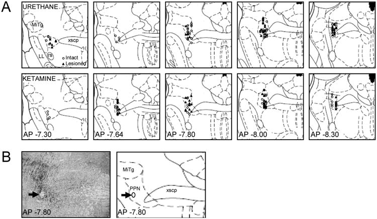
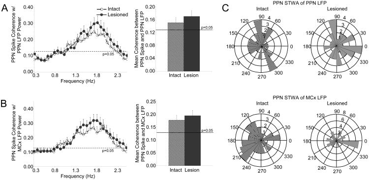
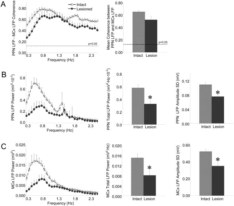
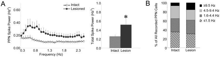
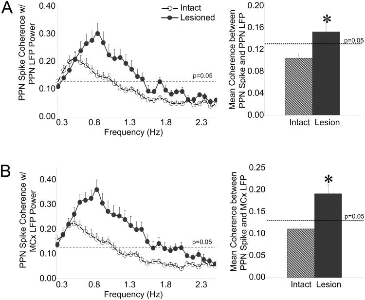
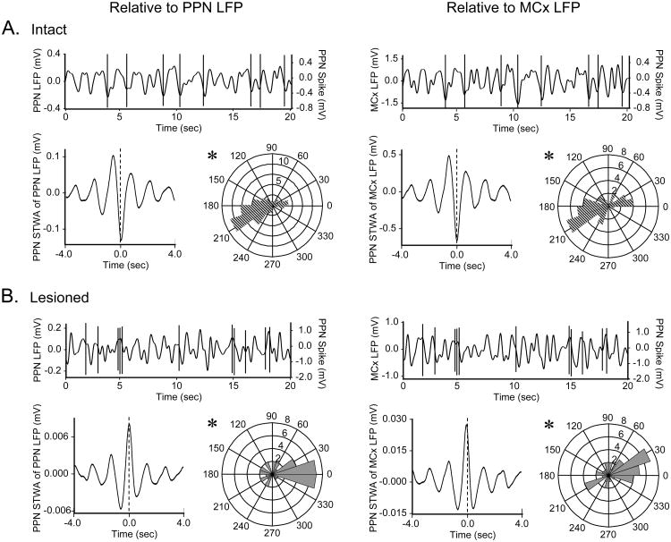
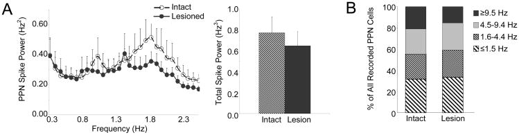
The pedunculopontine nucleus (PPN) is a new deep brain stimulation (DBS) target for Parkinson's disease (PD), but little is known about PPN firing pattern alterations in PD. The anesthetized rat is a useful model for investigating the effects of dopamine loss on the transmission of oscillatory cortical activity through basal ganglia structures. After dopamine loss, synchronous oscillatory activity emerges in the subthalamic nucleus and substantia nigra pars reticulata in phase with cortical slow oscillations. To investigate the impact of dopamine cell lesion-induced changes in basal ganglia output on activity in the PPN, this study examines PPN spike timing with reference to motor cortex (MCx) local field potential (LFP) activity in urethane- or ketamine-anesthetized rats. Seven to ten days after unilateral 6-hydroxydopamine lesion of the medial forebrain bundle, spectral power in PPN spike trains and coherence between PPN spiking and PPN LFP activity increased in the approximately 1 Hz range in urethane-anesthetized rats. PPN spike timing also changed from firing predominantly in phase with MCx slow oscillations in the intact urethane-anesthetized rat to firing predominantly antiphase to MCx oscillations in the hemi-parkinsonian rat. These changes were not observed in the ketamine-anesthetized preparation. These observations suggest that dopamine loss alters PPN spike timing by increasing inhibitory oscillatory input to the PPN from basal ganglia output nuclei, a phenomenon that may be relevant to motor dysfunction and PPN DBS efficacy in PD patients.
脚桥核(PPN)是帕金森病(PD)深部脑刺激(DBS)的一个新靶点,但关于PD中PPN放电模式的改变知之甚少。麻醉大鼠是研究多巴胺缺失对振荡性皮质活动通过基底神经节结构传递的影响的有用模型。多巴胺缺失后,丘脑底核和黑质网状部会出现与皮质慢振荡同步的振荡活动。为了研究多巴胺细胞损伤引起的基底神经节输出变化对PPN活动的影响,本研究在乌拉坦或氯胺酮麻醉的大鼠中,参照运动皮质(MCx)局部场电位(LFP)活动来检测PPN的尖峰时间。在单侧内侧前脑束6-羟基多巴胺损伤7至10天后,乌拉坦麻醉大鼠的PPN尖峰序列中的频谱功率以及PPN尖峰与PPN LFP活动之间的相干性在约1Hz范围内增加。PPN尖峰时间也从完整的乌拉坦麻醉大鼠中主要与MCx慢振荡同相放电转变为半帕金森病大鼠中主要与MCx振荡反相放电。在氯胺酮麻醉的实验准备中未观察到这些变化。这些观察结果表明,多巴胺缺失通过增加来自基底神经节输出核的对PPN的抑制性振荡输入来改变PPN尖峰时间,这一现象可能与PD患者的运动功能障碍和PPN DBS疗效相关。