• 文献检索
  • 文档翻译
  • 深度研究
  • 学术资讯
  • Suppr Zotero 插件Zotero 插件
  • 邀请有礼
  • 套餐&价格
  • 历史记录
应用&插件
Suppr Zotero 插件Zotero 插件浏览器插件Mac 客户端Windows 客户端微信小程序
定价
高级版会员购买积分包购买API积分包
服务
文献检索文档翻译深度研究API 文档MCP 服务
关于我们
关于 Suppr公司介绍联系我们用户协议隐私条款
关注我们

Suppr 超能文献

核心技术专利:CN118964589B侵权必究
粤ICP备2023148730 号-1Suppr @ 2026

文献检索

告别复杂PubMed语法,用中文像聊天一样搜索,搜遍4000万医学文献。AI智能推荐,让科研检索更轻松。

立即免费搜索

文件翻译

保留排版,准确专业,支持PDF/Word/PPT等文件格式,支持 12+语言互译。

免费翻译文档

深度研究

AI帮你快速写综述,25分钟生成高质量综述,智能提取关键信息,辅助科研写作。

立即免费体验

大鼠脑出血的颞部磁共振成像评估

Temporal MRI assessment of intracerebral hemorrhage in rats.

作者信息

Knight Robert A, Han Yuxia, Nagaraja Tavarekere N, Whitton Polly, Ding Jennifer, Chopp Michael, Seyfried Donald M

机构信息

Henry Ford Hospital, Department of Neurology-NMR Research, 2799 West Grand Blvd, Detroit, MI 48202, USA.

出版信息

Stroke. 2008 Sep;39(9):2596-602. doi: 10.1161/STROKEAHA.107.506683. Epub 2008 Jul 17.

DOI:10.1161/STROKEAHA.107.506683
PMID:18635862
原文链接:https://pmc.ncbi.nlm.nih.gov/articles/PMC3980853/
Abstract

BACKGROUND AND PURPOSE

MRI was used to evaluate the effects of experimental intracerebral hemorrhage (ICH) on brain tissue injury and recovery.

METHODS

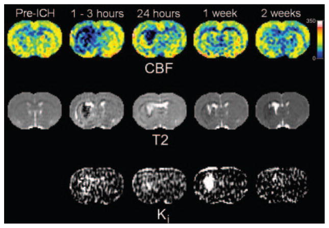
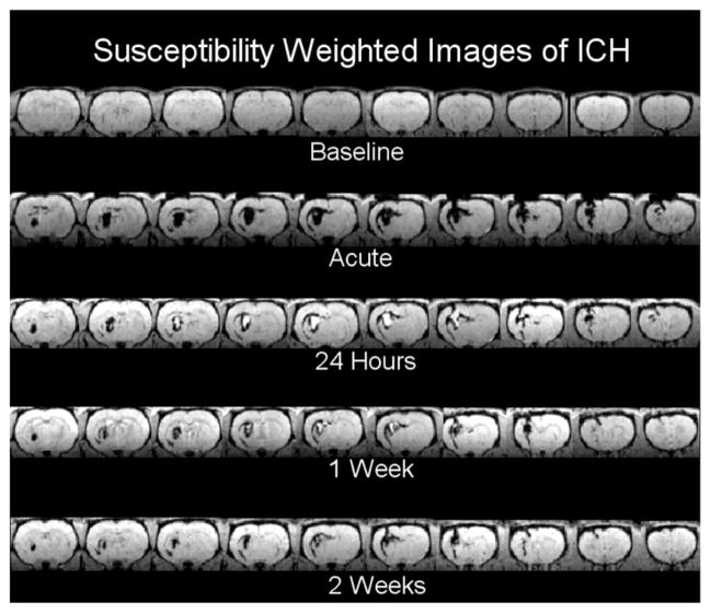
Primary ICH was induced in rats (n=6) by direct infusion of autologous blood into the striatum. The evolution of ICH damage was assessed by MRI estimates of T(2) and T(1sat) relaxation times, cerebral blood flow, vascular permeability, and susceptibility-weighted imaging before surgery (baseline) and at 2 hours and 1, 7, and 14 days post-ICH. Behavioral testing was done before and at 1, 7, and 14 days post-ICH. Animals were euthanized for histology at 14 days.

RESULTS

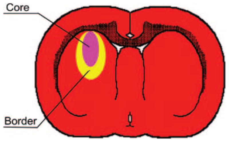
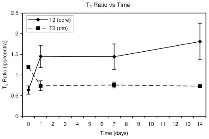
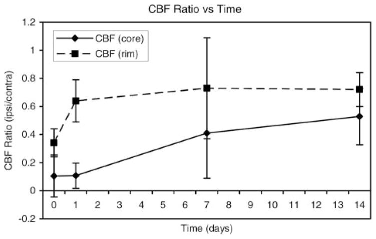
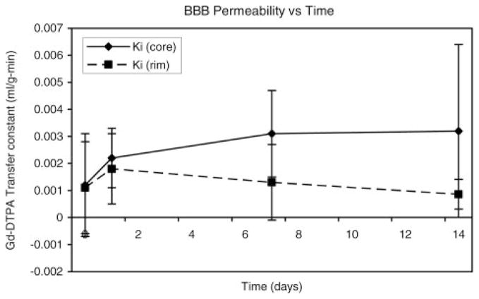
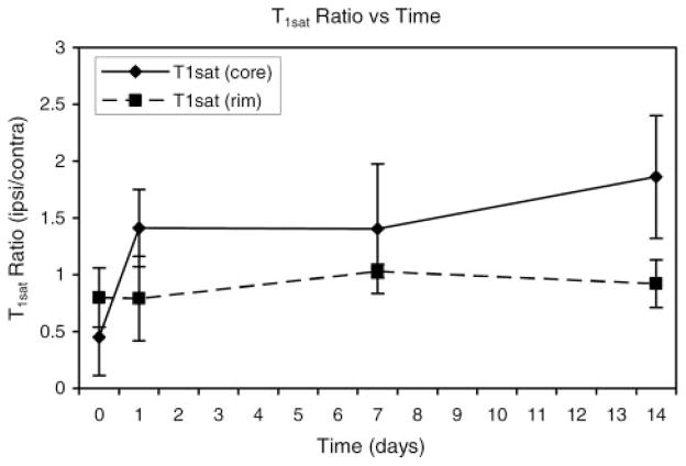
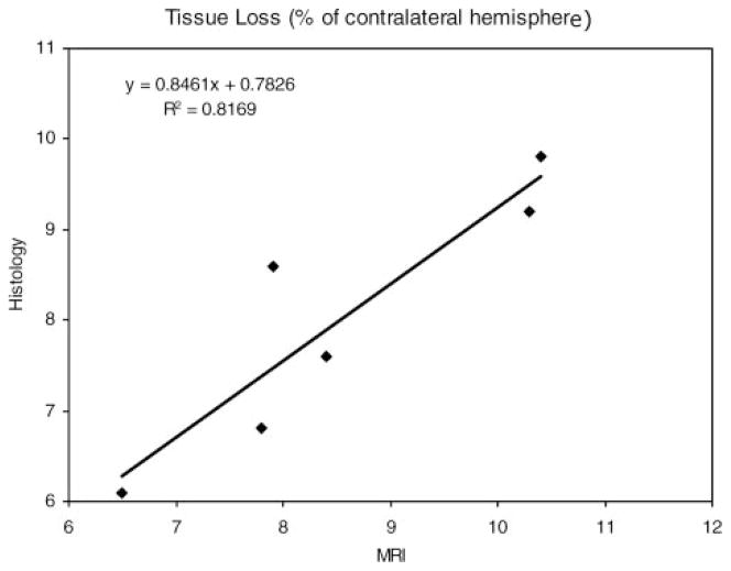
The MRI appearance of the hemorrhage and surrounding regions changed in a consistent manner over time. Two primary regions of interest were identified based on T(2) values. These included a core, corresponding to the bulk of the hemorrhage, and an adjacent rim; both varied with time. The core was associated with significantly lower cerebral blood flow values at all post-ICH time points, whereas cerebral blood flow varied in the rim. Increases in vascular permeability were noted at 1, 7, and 14 days. Changes in T(1sat) were similar to those of T(2). MRI and histological estimates of tissue loss were well correlated and showed approximately 9% hemispheric tissue loss.

CONCLUSIONS

Although the cerebral blood flow changes observed with this ICH model may not exactly mimic the clinical situation, our results suggest that the evolution of ICH injury can be accurately characterized with MRI. These methods may be useful to evaluate therapeutic interventions after experimental ICH and eventually in humans.

摘要

背景与目的

采用磁共振成像(MRI)评估实验性脑出血(ICH)对脑组织损伤及恢复的影响。

方法

通过将自体血直接注入大鼠(n = 6)纹状体诱导原发性ICH。在手术前(基线)以及ICH后2小时、1天、7天和14天,通过MRI评估T(2)和T(1sat)弛豫时间、脑血流量、血管通透性以及磁敏感加权成像,以评估ICH损伤的演变。在ICH前以及ICH后1天、7天和14天进行行为测试。在14天时对动物实施安乐死以进行组织学检查。

结果

出血及周围区域的MRI表现随时间呈一致变化。基于T(2)值确定了两个主要感兴趣区域。其中包括一个核心区,对应大部分出血,以及一个相邻的边缘区;两者均随时间变化。核心区在所有ICH后时间点的脑血流量值均显著较低,而边缘区的脑血流量有所变化。在1天、7天和14天时观察到血管通透性增加。T(1sat)的变化与T(2)相似。MRI与组织损失的组织学评估相关性良好,显示半球组织损失约9%。

结论

尽管此ICH模型观察到的脑血流量变化可能无法完全模拟临床情况,但我们的结果表明,MRI能够准确表征ICH损伤的演变。这些方法可能有助于评估实验性ICH后的治疗干预措施,并最终应用于人类。

https://cdn.ncbi.nlm.nih.gov/pmc/blobs/714e/3980853/1498923491f0/nihms567985f9.jpg
https://cdn.ncbi.nlm.nih.gov/pmc/blobs/714e/3980853/0e7a664ca647/nihms567985f1.jpg
https://cdn.ncbi.nlm.nih.gov/pmc/blobs/714e/3980853/c691fb2ea771/nihms567985f2.jpg
https://cdn.ncbi.nlm.nih.gov/pmc/blobs/714e/3980853/4c7aa69ec150/nihms567985f3.jpg
https://cdn.ncbi.nlm.nih.gov/pmc/blobs/714e/3980853/6e3386819bda/nihms567985f4.jpg
https://cdn.ncbi.nlm.nih.gov/pmc/blobs/714e/3980853/73d545723f45/nihms567985f5.jpg
https://cdn.ncbi.nlm.nih.gov/pmc/blobs/714e/3980853/c7e80ff92802/nihms567985f6.jpg
https://cdn.ncbi.nlm.nih.gov/pmc/blobs/714e/3980853/4f33d26b013b/nihms567985f7.jpg
https://cdn.ncbi.nlm.nih.gov/pmc/blobs/714e/3980853/912114e4640c/nihms567985f8.jpg
https://cdn.ncbi.nlm.nih.gov/pmc/blobs/714e/3980853/1498923491f0/nihms567985f9.jpg

相似文献

1
Temporal MRI assessment of intracerebral hemorrhage in rats.大鼠脑出血的颞部磁共振成像评估
Stroke. 2008 Sep;39(9):2596-602. doi: 10.1161/STROKEAHA.107.506683. Epub 2008 Jul 17.
2
Vascular recovery promoted by atorvastatin and simvastatin after experimental intracerebral hemorrhage: magnetic resonance imaging and histological study.阿托伐他汀和辛伐他汀促进实验性脑出血后血管恢复的磁共振成像和组织学研究。
J Neurosurg. 2011 Apr;114(4):1135-42. doi: 10.3171/2010.7.JNS10163. Epub 2010 Aug 20.
3
Experimental intracerebral hemorrhage: relationship between brain edema, blood flow, and blood-brain barrier permeability in rats.实验性脑出血:大鼠脑水肿、血流与血脑屏障通透性之间的关系
J Neurosurg. 1994 Jul;81(1):93-102. doi: 10.3171/jns.1994.81.1.0093.
4
Correlation between serum IL-1beta levels and cerebral edema extent in a hypertensive intracerebral hemorrhage rat model.高血压脑出血大鼠模型中血清白细胞介素-1β水平与脑水肿程度的相关性
Neurol Res. 2014 Feb;36(2):170-5. doi: 10.1179/1743132813Y.0000000292. Epub 2013 Dec 19.
5
Simvastatin and atorvastatin improve neurological outcome after experimental intracerebral hemorrhage.辛伐他汀和阿托伐他汀可改善实验性脑出血后的神经功能结局。
Stroke. 2009 Oct;40(10):3384-9. doi: 10.1161/STROKEAHA.108.544395. Epub 2009 Jul 30.
6
Glial-associated changes in the cerebral cortex after collagenase-induced intracerebral hemorrhage in the rat striatum.胶原酶诱导大鼠纹状体脑出血后脑皮质胶质相关变化。
Brain Res Bull. 2017 Sep;134:55-62. doi: 10.1016/j.brainresbull.2017.07.002. Epub 2017 Jul 10.
7
Prediction of impending hemorrhagic transformation in ischemic stroke using magnetic resonance imaging in rats.利用磁共振成像预测大鼠缺血性卒中即将发生的出血性转化
Stroke. 1998 Jan;29(1):144-51. doi: 10.1161/01.str.29.1.144.
8
Comparison of cerebral blood flow and injury following intracerebral and subdural hematoma in the rat.大鼠脑内和硬膜下血肿后脑血流量及损伤的比较。
Brain Res. 1999 May 22;829(1-2):125-33. doi: 10.1016/s0006-8993(99)01378-5.
9
Experimental intracerebral hematoma in the rat: characterization by sequential magnetic resonance imaging, behavior, and histopathology. Effect of albumin therapy.大鼠实验性脑内血肿:通过序列磁共振成像、行为学和组织病理学进行特征描述。白蛋白治疗的效果。
Brain Res. 2007 Jul 9;1157:146-55. doi: 10.1016/j.brainres.2007.04.077. Epub 2007 May 8.
10
Lobar intracerebral hemorrhage model in pigs: rapid edema development in perihematomal white matter.猪脑叶脑出血模型:血肿周围白质快速出现水肿
Stroke. 1996 Mar;27(3):490-7. doi: 10.1161/01.str.27.3.490.

引用本文的文献

1
Multimodal and serial MRI monitors brain peri-hematomal injury and repair mechanisms after experimental intracerebral hemorrhage.多模态和序列磁共振成像监测实验性脑出血后脑血肿周围损伤及修复机制。
J Cereb Blood Flow Metab. 2025 Jan;45(1):140-152. doi: 10.1177/0271678X241270198. Epub 2024 Aug 7.
2
Enhanced liver X receptor signalling reduces brain injury and promotes tissue regeneration following experimental intracerebral haemorrhage: roles of microglia/macrophages.增强肝 X 受体信号可减轻实验性脑出血后的脑损伤并促进组织再生:小胶质细胞/巨噬细胞的作用。
Stroke Vasc Neurol. 2023 Dec 29;8(6):486-502. doi: 10.1136/svn-2023-002331.
3
Experimental animal models and evaluation techniques in intracerebral hemorrhage.脑出血的实验动物模型及评估技术
Tzu Chi Med J. 2022 Jul 26;35(1):1-10. doi: 10.4103/tcmj.tcmj_119_22. eCollection 2023 Jan-Mar.
4
Photothrombotic Middle Cerebral Artery Occlusion in Mice: A Novel Model of Ischemic Stroke.光血栓性大脑中动脉阻塞在小鼠中的应用:一种新的缺血性脑卒中模型。
eNeuro. 2023 Feb 8;10(2). doi: 10.1523/ENEURO.0244-22.2022. Print 2023 Feb.
5
Intracerebral Hemorrhage-Induced Cognitive Impairment in Rats Is Associated With Brain Atrophy, Hypometabolism, and Network Dysconnectivity.大鼠脑出血所致认知障碍与脑萎缩、代谢减退及网络连接障碍有关。
Front Neurosci. 2022 Jun 30;16:882996. doi: 10.3389/fnins.2022.882996. eCollection 2022.
6
Early brainstem hemorrhage progression: multi-sequence magnetic resonance imaging and histopathology.早期脑干出血进展:多序列磁共振成像与组织病理学
Neural Regen Res. 2023 Jan;18(1):170-175. doi: 10.4103/1673-5374.344838.
7
A Magnetic Resonance-Relaxometry-Based Technique to Identify Blood Products in Brain Parenchyma: An Experimental Study on a Rabbit Model.一种基于磁共振弛豫测量法的技术用于识别脑实质中的血液制品:在兔模型上的实验研究
Front Vet Sci. 2022 May 31;9:802272. doi: 10.3389/fvets.2022.802272. eCollection 2022.
8
B-Mode Ultrasound, a Reliable Tool for Monitoring Experimental Intracerebral Hemorrhage.B 型超声:监测实验性脑出血的可靠工具
Front Neurol. 2021 Dec 23;12:771402. doi: 10.3389/fneur.2021.771402. eCollection 2021.
9
Translational Stroke Research Review: Using the Mouse to Model Human Futile Recanalization and Reperfusion Injury in Ischemic Brain Tissue.转化卒中研究综述:利用小鼠模型模拟缺血性脑组织中无效再通和再灌注损伤。
Cells. 2021 Nov 25;10(12):3308. doi: 10.3390/cells10123308.
10
Simvastatin-Ezetimibe enhances growth factor expression and attenuates neuron loss in the hippocampus in a model of intracerebral hemorrhage.辛伐他汀-依折麦布增强脑出血模型中海马生长因子表达并减轻神经元丢失。
Fundam Clin Pharmacol. 2021 Aug;35(4):634-644. doi: 10.1111/fcp.12635. Epub 2021 Jan 19.

本文引用的文献

1
Acute hypertension in intracerebral hemorrhage: pathophysiology and treatment.脑出血中的急性高血压:病理生理学与治疗
J Neurol Sci. 2007 Oct 15;261(1-2):74-9. doi: 10.1016/j.jns.2007.04.036. Epub 2007 Jun 5.
2
Experimental intracerebral hematoma in the rat: characterization by sequential magnetic resonance imaging, behavior, and histopathology. Effect of albumin therapy.大鼠实验性脑内血肿:通过序列磁共振成像、行为学和组织病理学进行特征描述。白蛋白治疗的效果。
Brain Res. 2007 Jul 9;1157:146-55. doi: 10.1016/j.brainres.2007.04.077. Epub 2007 May 8.
3
Guidelines for the management of spontaneous intracerebral hemorrhage in adults: 2007 update: a guideline from the American Heart Association/American Stroke Association Stroke Council, High Blood Pressure Research Council, and the Quality of Care and Outcomes in Research Interdisciplinary Working Group.成人自发性脑出血管理指南:2007年更新版:美国心脏协会/美国卒中协会卒中委员会、高血压研究委员会以及研究中的医疗质量与结局跨学科工作组制定的指南
Stroke. 2007 Jun;38(6):2001-23. doi: 10.1161/STROKEAHA.107.183689. Epub 2007 May 3.
4
Quantitative perihematomal blood flow in spontaneous intracerebral hemorrhage predicts in-hospital functional outcome.自发性脑出血周围血肿定量血流可预测住院期间的功能结局。
Stroke. 2007 Feb;38(2):319-24. doi: 10.1161/01.STR.0000254492.35504.db. Epub 2006 Dec 28.
5
Perfusion-weighted magnetic resonance imaging in acute intracerebral hemorrhage at baseline and during the 1st and 2nd week: a longitudinal study.急性脑出血患者基线期、第1周和第2周的灌注加权磁共振成像:一项纵向研究
Cerebrovasc Dis. 2007;23(1):6-13. doi: 10.1159/000095752. Epub 2006 Sep 12.
6
Perihematomal mitochondrial dysfunction after intracerebral hemorrhage.脑出血后血肿周围线粒体功能障碍
Stroke. 2006 Oct;37(10):2457-62. doi: 10.1161/01.STR.0000240674.99945.4e. Epub 2006 Sep 7.
7
Mast cell blocking reduces brain edema and hematoma volume and improves outcome after experimental intracerebral hemorrhage.肥大细胞阻断可减轻实验性脑出血后的脑水肿和血肿体积,并改善预后。
J Cereb Blood Flow Metab. 2007 Apr;27(4):795-802. doi: 10.1038/sj.jcbfm.9600387. Epub 2006 Aug 16.
8
Effects of intravenous administration of human bone marrow stromal cells after intracerebral hemorrhage in rats.大鼠脑出血后静脉注射人骨髓基质细胞的效果
J Neurosurg. 2006 Feb;104(2):313-8. doi: 10.3171/jns.2006.104.2.313.
9
Acute blood-brain barrier opening in experimentally induced focal cerebral ischemia is preferentially identified by quantitative magnetization transfer imaging.在实验性诱导的局灶性脑缺血中,急性血脑屏障开放可通过定量磁化传递成像优先识别。
Magn Reson Med. 2005 Oct;54(4):822-32. doi: 10.1002/mrm.20630.
10
Quantitation and localization of blood-to-brain influx by magnetic resonance imaging and quantitative autoradiography in a model of transient focal ischemia.在短暂性局灶性缺血模型中,通过磁共振成像和定量放射自显影对血脑屏障通透性进行定量和定位分析。
Magn Reson Med. 2005 Oct;54(4):813-21. doi: 10.1002/mrm.20629.