Departments of Neurosurgery, Henry Ford Hospital, Detroit, Michigan 48202, USA.
J Neurosurg. 2011 Apr;114(4):1135-42. doi: 10.3171/2010.7.JNS10163. Epub 2010 Aug 20.
Longitudinal multiparametric MR imaging and histological studies were performed on simvastatin- or atorvastatin-treated rats to evaluate vascular repair mechanisms after experimental intracerebral hemorrhage (ICH).
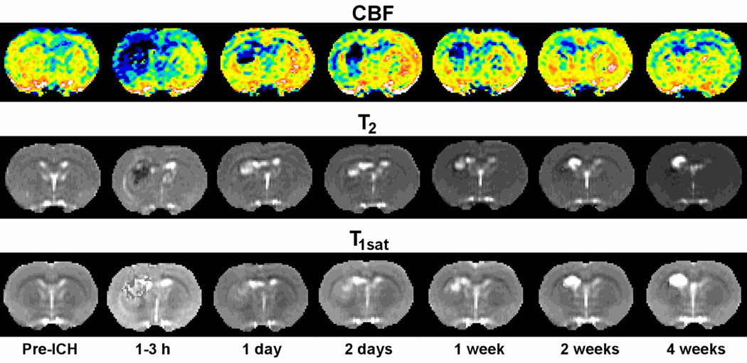
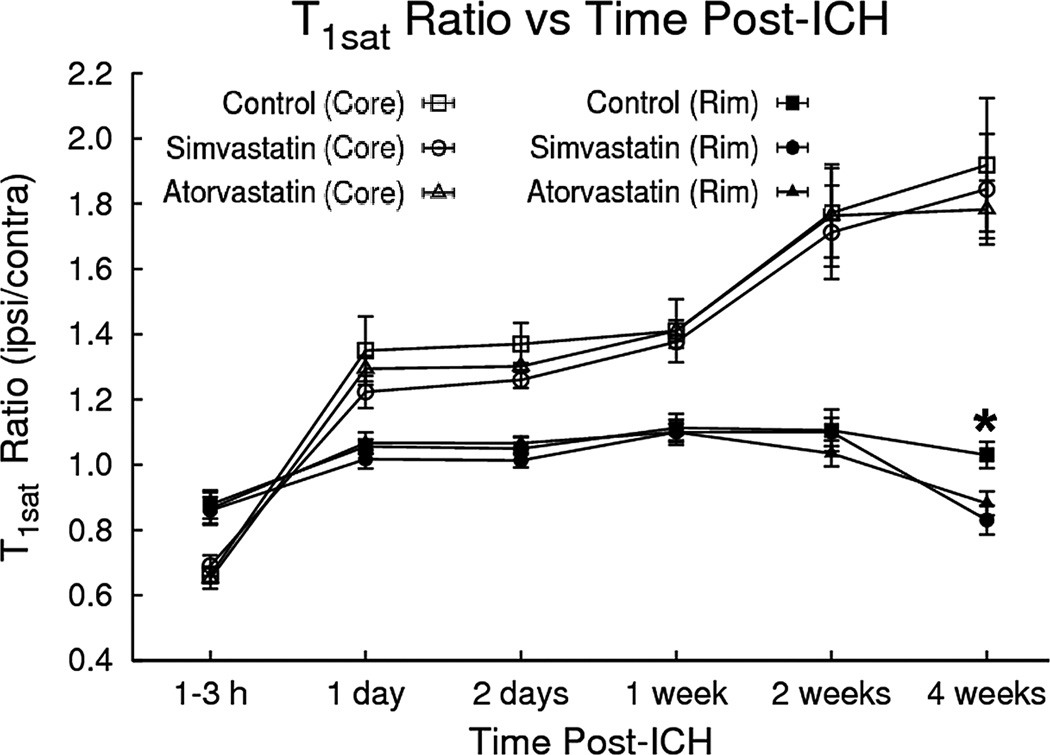
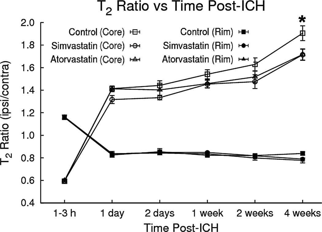
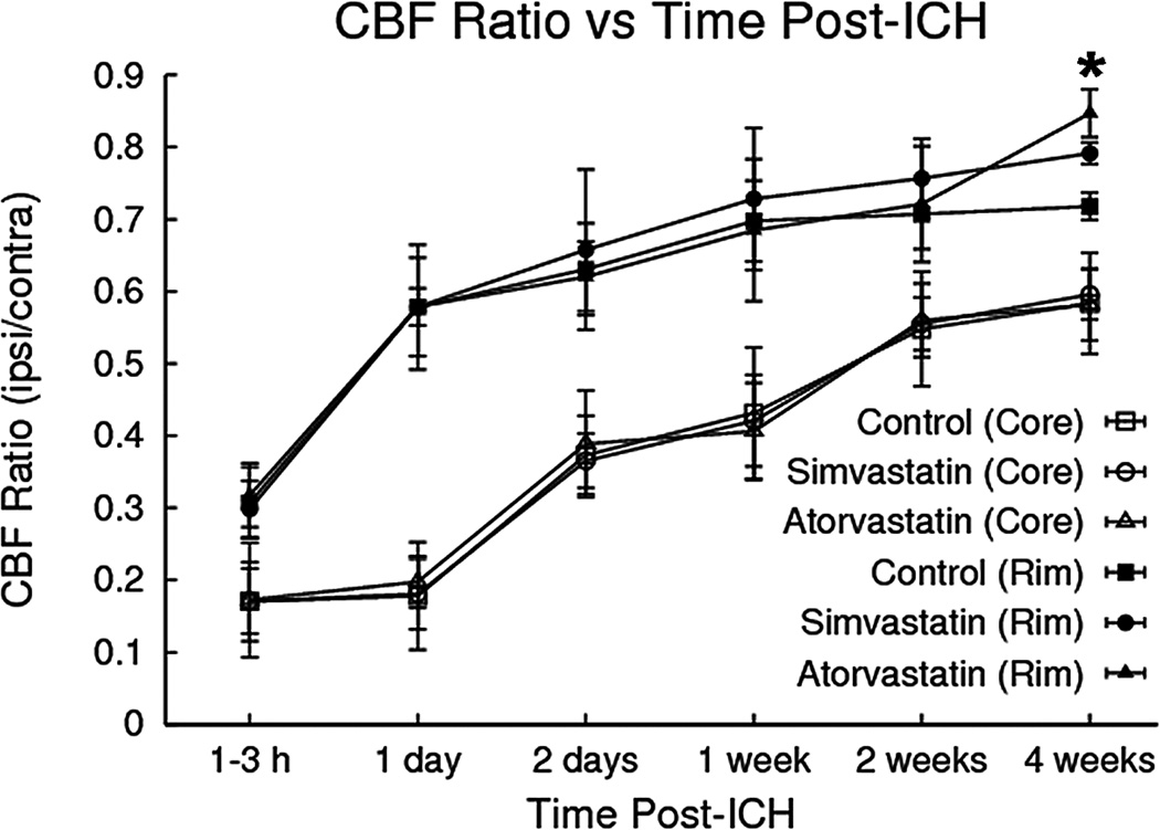
Primary ICH was induced in adult Wistar rats by direct infusion of 100 μl of autologous blood into the striatal region adjacent to the subventricular zone. Atorvastatin (2 mg/kg), simvastatin (2 mg/kg), or phosphate-buffered saline was given orally at 24 hours post-ICH and continued daily for 7 days. The temporal evolution of ICH in each group was assessed by MR imaging measurements of T2, T1(sat), and cerebral blood flow in brain areas corresponding to the bulk of the hemorrhage (core) and edematous border (rim). Rats were killed after the final MR imaging examination at 28 days, and histological studies were performed. A small group of sham-operated animals was also studied. Neurobehavioral testing was performed in all animals. Analysis of variance methods were used to compare results from the treatment and control groups, with significance inferred at p ≤ 0.05.
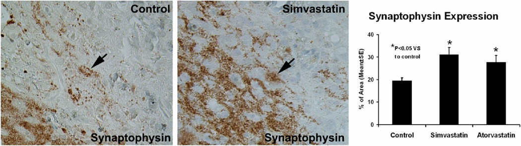
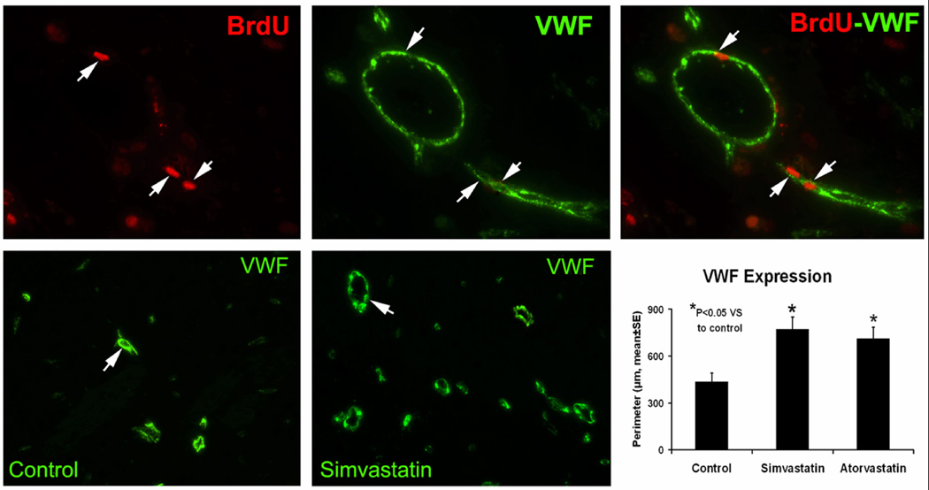
Using histological indices, animals treated with simvastatin and atorvastatin had significantly increased angiogenesis and synaptogenesis in the hematoma rim compared with the control group (p ≤ 0.05). The statin-treated animals exhibited significantly increased cerebral blood flow in the hematoma rim at 4 weeks, while blood-brain barrier permeability (T1(sat)) and edema (T2) in the corresponding regions were reduced. Both statin-treated groups showed significant neurological improvement from 2 weeks post-ICH onward.
The results of the present study demonstrate that simvastatin and atorvastatin significantly improve the recovery of rats from ICH, possibly via angiogenesis and synaptic plasticity. In addition, in vivo multiparametric MR imaging measurements over time can be effectively applied to the experimental ICH model for longitudinal assessment of the therapeutic intervention.
对辛伐他汀或阿托伐他汀治疗的大鼠进行纵向多参数磁共振成像和组织学研究,以评估实验性脑出血(ICH)后血管修复机制。
通过将 100μl 自体血液直接输注到靠近侧脑室区域的纹状体区域,在成年 Wistar 大鼠中诱导原发性 ICH。在 ICH 后 24 小时给予阿托伐他汀(2mg/kg)、辛伐他汀(2mg/kg)或磷酸盐缓冲盐水口服,每天一次,持续 7 天。通过磁共振成像测量 T2、T1(sat)和脑血流,评估每组 ICH 的时间演变,这些测量值来自与血肿大部分相对应的脑区(核心)和水肿边界(边缘)。在 28 天的最后一次磁共振成像检查后,大鼠被处死,并进行组织学研究。还研究了一小部分假手术动物。所有动物均进行神经行为测试。使用方差分析方法比较治疗组和对照组的结果,显著性推断 p≤0.05。
使用组织学指标,与对照组相比,辛伐他汀和阿托伐他汀治疗的动物在血肿边缘的血管生成和突触发生明显增加(p≤0.05)。他汀类治疗动物在 4 周时在血肿边缘显示出明显增加的脑血流,而相应区域的血脑屏障通透性(T1(sat))和水肿(T2)降低。从 ICH 后 2 周开始,两组他汀类治疗动物均表现出明显的神经功能改善。
本研究结果表明,辛伐他汀和阿托伐他汀可显著改善 ICH 大鼠的恢复,可能通过血管生成和突触可塑性。此外,随着时间的推移,体内多参数磁共振成像测量可有效地应用于实验性 ICH 模型,用于对治疗干预的纵向评估。