Karki Kishor, Knight Robert A, Han Yuxia, Yang Dongmei, Zhang Jianfeng, Ledbetter Karyn A, Chopp Michael, Seyfried Donald M
Department of Neurology, Henry Ford Hospital, Detroit, MI 48202, USA.
Stroke. 2009 Oct;40(10):3384-9. doi: 10.1161/STROKEAHA.108.544395. Epub 2009 Jul 30.
This study investigates the effects of statin treatment on experimental intracerebral hemorrhage (ICH) using behavioral, histological, and MRI measures of recovery.
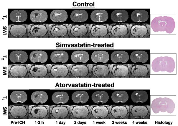
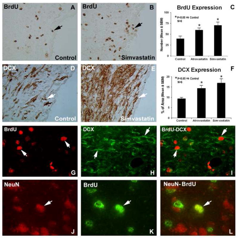
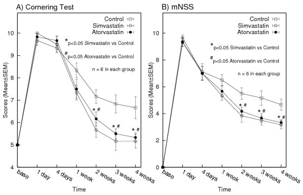
Primary ICH was induced in rats. Simvastatin (2 mg/kg), atorvastatin (2 mg/kg), or phosphate-buffered saline (n=6 per group) was given daily for 1 week. MRI studies were performed 2 to 3 days before ICH, and at 1 to 2 hours and 1, 2, 7, 14, and 28 days after ICH. The ICH evolution was assessed via hematoma volume measurements using susceptibility-weighted imaging (SWI) and tissue loss using T2 maps and hematoxylin and eosin (H&E) histology. Neurobehavioral tests were done before ICH and at various time points post-ICH. Additional histological measures were performed with doublecortin neuronal nuclei and bromodeoxyuridine stainings.
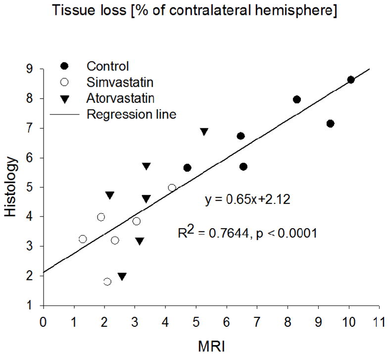
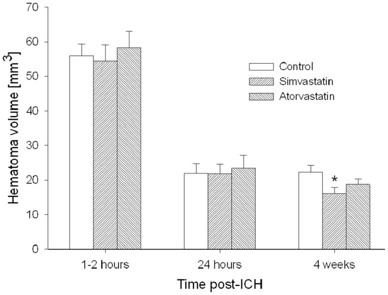
Initial ICH volumes determined by SWI were similar across all groups. Simvastatin significantly reduced hematoma volume at 4 weeks (P=0.002 versus control with acute volumes as baseline), whereas that for atorvastatin was marginal (P=0.09). MRI estimates of tissue loss (% of contralateral hemisphere) for treated rats were significantly lower (P=0.0003 and 0.001, respectively) than for control at 4 weeks. Similar results were obtained for H&E histology (P=0.0003 and 0.02, respectively). Tissue loss estimates between MRI and histology were well correlated (R2=0.764, P<0.0001). Significant improvement in neurological function was seen 2 to 4 weeks post-ICH with increased neurogenesis observed.
Simvastatin and atorvastatin significantly improved neurological recovery, decreased tissue loss, and increased neurogenesis when administered for 1 week after ICH.
本研究使用行为学、组织学和MRI恢复指标,调查他汀类药物治疗对实验性脑出血(ICH)的影响。
在大鼠中诱导原发性ICH。每天给予辛伐他汀(2mg/kg)、阿托伐他汀(2mg/kg)或磷酸盐缓冲盐水(每组n = 6),持续1周。在ICH前2至3天以及ICH后1至2小时、1天、2天、7天、14天和28天进行MRI研究。通过使用磁敏感加权成像(SWI)测量血肿体积以及使用T2图和苏木精-伊红(H&E)组织学评估组织损失来评估ICH演变。在ICH前和ICH后的各个时间点进行神经行为测试。使用双皮质素神经元核和溴脱氧尿苷染色进行额外的组织学测量。
通过SWI确定的初始ICH体积在所有组中相似。辛伐他汀在4周时显著降低了血肿体积(与以急性体积为基线的对照组相比,P = 0.002),而阿托伐他汀的作用不明显(P = 0.09)。治疗大鼠的MRI组织损失估计值(对侧半球的百分比)在4周时显著低于对照组(分别为P = 0.0003和0.001)。H&E组织学也得到了类似的结果(分别为P = 0.0003和0.02)。MRI和组织学之间的组织损失估计值具有良好的相关性(R2 = 0.764,P < 0.0001)。ICH后2至4周观察到神经功能有显著改善,同时神经发生增加。
ICH后给予辛伐他汀和阿托伐他汀1周,可显著改善神经功能恢复,减少组织损失,并增加神经发生。