Schelhaas Mario, Ewers Helge, Rajamäki Minna-Liisa, Day Patricia M, Schiller John T, Helenius Ari
Institute of Biochemistry, ETH Zurich, Zurich, Switzerland.
PLoS Pathog. 2008 Sep 5;4(9):e1000148. doi: 10.1371/journal.ppat.1000148.
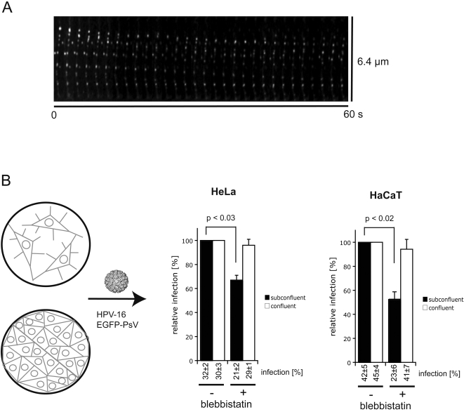
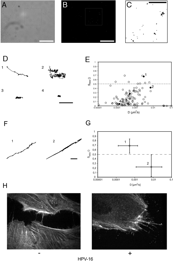
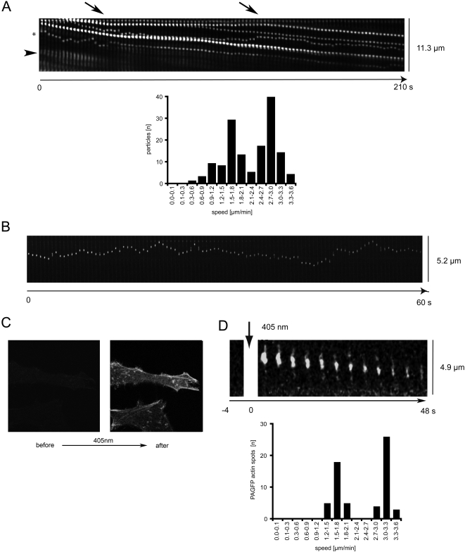
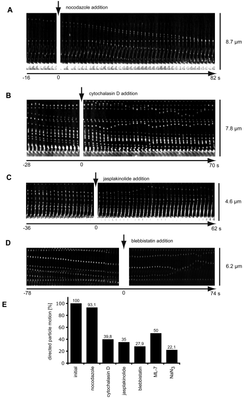
The lateral mobility of individual, incoming human papillomavirus type 16 pseudoviruses (PsV) bound to live HeLa cells was studied by single particle tracking using fluorescence video microscopy. The trajectories were computationally analyzed in terms of diffusion rate and mode of motion as described by the moment scaling spectrum. Four distinct modes of mobility were seen: confined movement in small zones (30-60 nm in diameter), confined movement with a slow drift, fast random motion with transient confinement, and linear, directed movement for long distances. The directed movement was most prominent on actin-rich cell protrusions such as filopodia or retraction fibres, where the rate was similar to that measured for actin retrograde flow. It was, moreover, sensitive to perturbants of actin retrograde flow such as cytochalasin D, jasplakinolide, and blebbistatin. We found that transport along actin protrusions significantly enhanced HPV-16 infection in sparse tissue culture, cells suggesting a role for in vivo infection of basal keratinocytes during wound healing.
利用荧光视频显微镜通过单粒子追踪研究了与活的HeLa细胞结合的单个传入人乳头瘤病毒16型假病毒(PsV)的侧向迁移率。按照矩量缩放谱所描述的,对轨迹在扩散速率和运动模式方面进行了计算分析。观察到四种不同的迁移模式:在小区域(直径30 - 60纳米)内的受限运动、伴有缓慢漂移的受限运动、具有瞬时受限的快速随机运动以及长距离的线性定向运动。定向运动在富含肌动蛋白的细胞突起如丝状伪足或收缩纤维上最为显著,其速率与肌动蛋白逆行流所测得的速率相似。此外,它对肌动蛋白逆行流的干扰剂如细胞松弛素D、茉莉酸甲酯和肌球蛋白II抑制剂敏感。我们发现,在稀疏组织培养细胞中,沿肌动蛋白突起的转运显著增强了HPV - 16感染,这表明在伤口愈合过程中对基底角质形成细胞的体内感染具有作用。