Luther Pradeep K, Bennett Pauline M, Knupp Carlo, Craig Roger, Padrón Raúl, Harris Samantha P, Patel Jitendrakumar, Moss Richard L
Molecular Medicine Section, National Heart and Lung Institute, Faculty of Medicine, Imperial College London, London SW72AZ, UK.
J Mol Biol. 2008 Dec 5;384(1):60-72. doi: 10.1016/j.jmb.2008.09.013. Epub 2008 Sep 16.
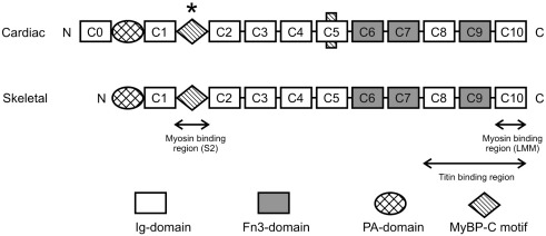
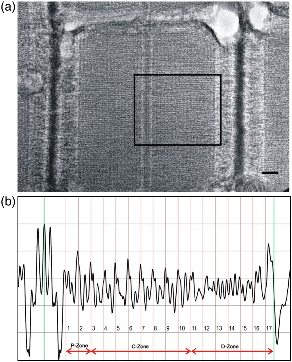
Myosin binding protein C (MyBP-C) is a component of the thick filament of striated muscle. The importance of this protein is revealed by recent evidence that mutations in the cardiac gene are a major cause of familial hypertrophic cardiomyopathy. Here we investigate the distribution of MyBP-C in the A-bands of cardiac and skeletal muscles and compare this to the A-band structure in cardiac muscle of MyBP-C-deficient mice. We have used a novel averaging technique to obtain the axial density distribution of A-bands in electron micrographs of well-preserved specimens. We show that cardiac and skeletal A-bands are very similar, with a length of 1.58+/-0.01 mum. In normal cardiac and skeletal muscle, the distributions are very similar, showing clearly the series of 11 prominent accessory protein stripes in each half of the A-band spaced axially at 43-nm intervals and starting at the edge of the bare zone. We show by antibody labelling that in cardiac muscle the distal nine stripes are the location of MyBP-C. These stripes are considerably suppressed in the knockout mouse hearts as expected. Myosin heads on the surface of the thick filament in relaxed muscle are thought to be arranged in a three-stranded quasi-helix with a mean 14.3-nm axial cross bridge spacing and a 43 nm helix repeat. Extra "forbidden" meridional reflections, at orders of 43 nm, in X-ray diffraction patterns of muscle have been interpreted as due to an axial perturbation of some levels of myosin heads. However, in the MyBP-C-deficient hearts these extra meridional reflections are weak or absent, suggesting that they are due to MyBP-C itself or to MyBP-C in combination with a head perturbation brought about by the presence of MyBP-C.
肌球蛋白结合蛋白C(MyBP-C)是横纹肌粗肌丝的一个组成部分。近期有证据表明,心脏基因的突变是家族性肥厚型心肌病的主要病因,这揭示了该蛋白的重要性。在此,我们研究了MyBP-C在心肌和骨骼肌A带中的分布,并将其与MyBP-C缺陷型小鼠心肌中的A带结构进行比较。我们使用了一种新颖的平均技术,以获取保存完好的标本电子显微照片中A带的轴向密度分布。我们发现,心肌和骨骼肌的A带非常相似,长度为1.58±0.01μm。在正常的心肌和骨骼肌中,分布非常相似,清晰地显示出A带每半部分有一系列11条突出的辅助蛋白条纹,轴向间隔为43nm,从裸区边缘开始。我们通过抗体标记表明,在心肌中,远端的九条条纹是MyBP-C的位置。正如预期的那样,在基因敲除小鼠的心脏中,这些条纹受到了显著抑制。在舒张状态下,粗肌丝表面的肌球蛋白头部被认为以三股准螺旋排列,平均轴向横桥间距为14.3nm,螺旋重复周期为43nm。肌肉X射线衍射图谱中43nm级别的额外“禁阻”子午反射被解释为是由于肌球蛋白头部某些水平的轴向扰动所致。然而,在MyBP-C缺陷型心脏中,这些额外的子午反射较弱或不存在,这表明它们是由MyBP-C本身或MyBP-C与MyBP-C的存在所引起的头部扰动共同导致的。