Valcárcel María, Arteta Beatriz, Jaureguibeitia Arrate, Lopategi Aritz, Martínez Iñigo, Mendoza Lorea, Muruzabal Francisco J, Salado Clarisa, Vidal-Vanaclocha Fernando
Dept, Cell Biology and Histology, Basque Country University School of Medicine & Dentistry, Bizkaia-48940, Spain.
J Transl Med. 2008 Oct 9;6:57. doi: 10.1186/1479-5876-6-57.
The recruitment of vascular stromal and endothelial cells is an early event occurring during cancer cell growth at premetastatic niches, but how the microenvironment created by the initial three-dimensional (3D) growth of cancer cells affects their angiogenesis-stimulating potential is unclear.
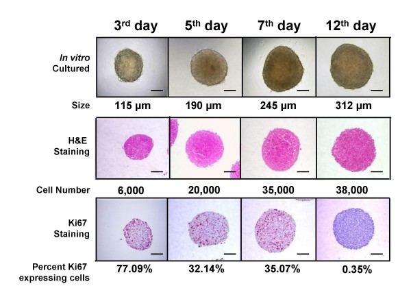
The proangiogenic profile of CT26 murine colorectal carcinoma cells was studied in seven-day cultured 3D-spheroids of <300 mum in diameter, produced by the hanging-drop method to mimic the microenvironment of avascular micrometastases prior to hypoxia occurrence.
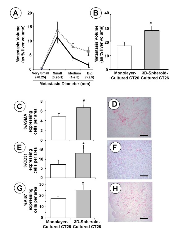
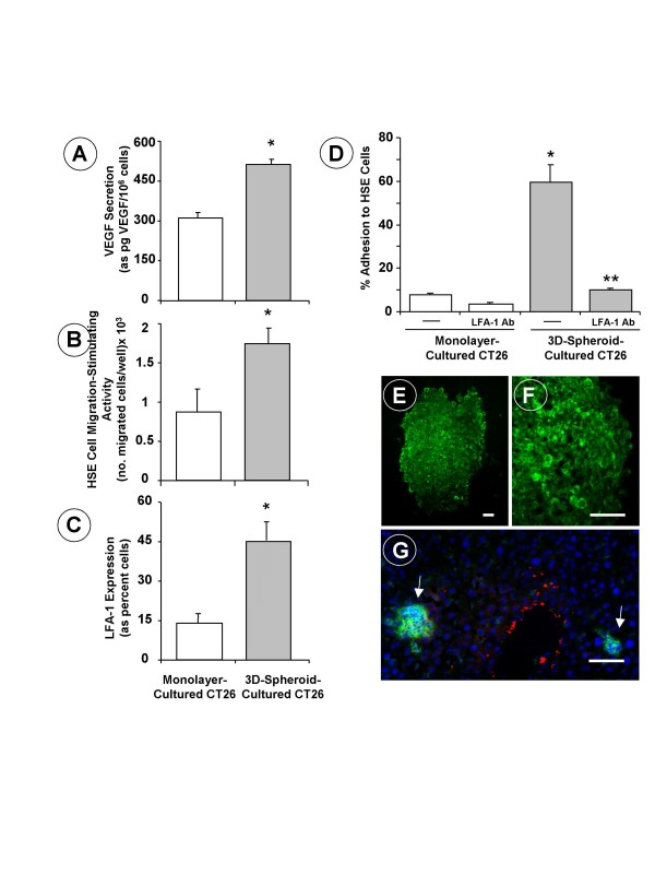
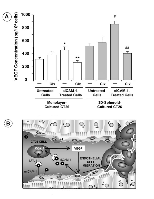
Spheroid-derived CT26 cells increased vascular endothelial growth factor (VEGF) secretion by 70%, which in turn increased the in vitro migration of primary cultured hepatic sinusoidal endothelium (HSE) cells by 2-fold. More importantly, spheroid-derived CT26 cells increased lymphocyte function associated antigen (LFA)-1-expressing cell fraction by 3-fold; and soluble intercellular adhesion molecule (ICAM)-1, given to spheroid-cultured CT26 cells, further increased VEGF secretion by 90%, via cyclooxygenase (COX)-2-dependent mechanism. Consistent with these findings, CT26 cancer cells significantly increased LFA-1 expression in non-hypoxic avascular micrometastases at their earliest inception within hepatic lobules in vivo; and angiogenesis also markedly increased in both subcutaneous tumors and hepatic metastases produced by spheroid-derived CT26 cells.
3D-growth per se enriched the proangiogenic phenotype of cancer cells growing as multicellular spheroids or as subclinical hepatic micrometastases. The contribution of integrin LFA-1 to VEGF secretion via COX-2 was a micro environmental-related mechanism leading to the pro-angiogenic activation of soluble ICAM-1-activated colorectal carcinoma cells. This mechanism may represent a new target for specific therapeutic strategies designed to block colorectal cancer cell growth at a subclinical micrometastatic stage within the liver.
血管基质细胞和内皮细胞的募集是癌细胞在转移前微环境中生长过程中发生的早期事件,但癌细胞最初的三维(3D)生长所形成的微环境如何影响其促血管生成潜力尚不清楚。
采用悬滴法培养直径小于300μm的7天龄3D球体,以模拟缺氧发生前无血管微转移灶的微环境,研究CT26小鼠结肠癌细胞的促血管生成特征。
球体来源的CT26细胞使血管内皮生长因子(VEGF)分泌增加70%,进而使原代培养的肝窦内皮(HSE)细胞的体外迁移增加2倍。更重要的是,球体来源的CT26细胞使表达淋巴细胞功能相关抗原(LFA)-1的细胞比例增加3倍;给予球体培养的CT26细胞可溶性细胞间黏附分子(ICAM)-1,通过环氧化酶(COX)-2依赖机制使VEGF分泌进一步增加90%。与这些发现一致,CT26癌细胞在体内肝小叶内最早出现的非缺氧无血管微转移灶中显著增加LFA-1表达;球体来源的CT26细胞产生的皮下肿瘤和肝转移灶中的血管生成也明显增加。
3D生长本身丰富了以多细胞球体或亚临床肝微转移灶形式生长的癌细胞的促血管生成表型。整合素LFA-1通过COX-2对VEGF分泌的作用是一种与微环境相关的机制,导致可溶性ICAM-1激活的结肠癌细胞发生促血管生成激活。这一机制可能代表了一种新的靶点,用于设计特定的治疗策略,以阻断肝内亚临床微转移阶段的结肠癌细胞生长。