Mello de Queiroz Fernanda, Ponte Cristiano G, Bonomo Adriana, Vianna-Jorge Rosane, Suarez-Kurtz Guilherme
Molekulare Biologie Neuronaler Signale, Max-Planck-Institut für Experimentelle Medizin, Hermann-Rein-Strasse 3, 37075 Göttingen, Germany.
BMC Immunol. 2008 Nov 3;9:63. doi: 10.1186/1471-2172-9-63.
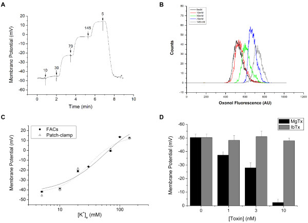
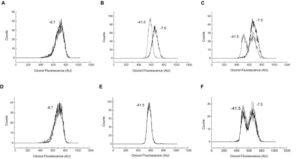
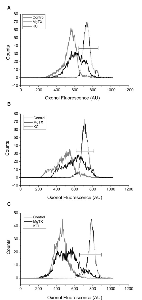
Ion channels are involved in the control of membrane potential (psi) in a variety of cells. The maintenance of psi in human T lymphocytes is essential for T-cell activation and was suggested to depend mostly on the voltage-gated Kv1.3 channel. Blockage of Kv1.3 inhibits cytokine production and lymphocyte proliferation in vitro and suppresses immune response in vivo. T lymphocytes are a heterogeneous cell population and the expression of Kv1.3 varies among cell subsets. Oxonol diBA-C4-(3) was used to determine psi by flow cytometry. The presence of distinct T cell subsets was evaluated by immunophenotyping techniques and the contribution of Kv1.3 channels for the maintenance of psi was investigated using selective blockers.
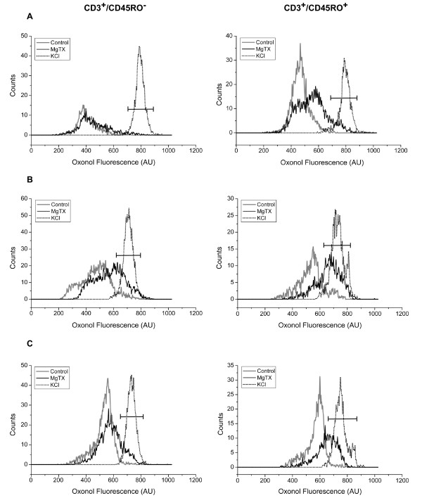
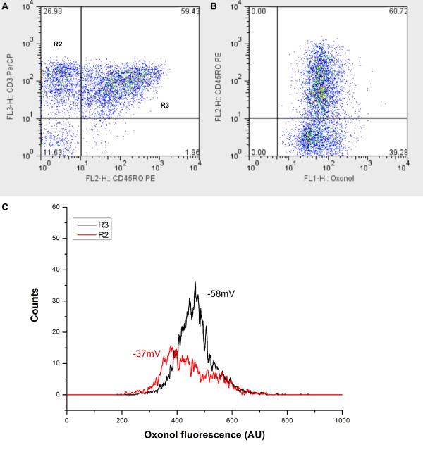
The distribution of psi in T lymphocytes varied among blood donors and did not always follow a unimodal pattern. T lymphocytes were divided into CD3+/CD45RO- and CD3+/CD45RO+ subsets, whose peak channel values of psi were -58 +/- 3.6 mV and -37 +/- 4.1 mV, respectively. MgTX (specific inhibitor of Kv1.3 channels) had no significant effect in the psi of CD3+/CD45RO- subsets but depolarized CD3+/CD45RO+ cells to -27 +/- 5.1 mV.
Combination of optical methods for determination of psi by flow cytometry with immuophenotyping techniques opens new possibilities for the study of ion channels in the biology of heterogeneous cell populations such as T lymphocyte subsets.
离子通道参与多种细胞中膜电位(ψ)的调控。人T淋巴细胞中ψ的维持对于T细胞活化至关重要,并且提示其主要依赖于电压门控的Kv1.3通道。阻断Kv1.3可抑制体外细胞因子的产生和淋巴细胞增殖,并在体内抑制免疫反应。T淋巴细胞是异质性细胞群体,Kv1.3的表达在细胞亚群间存在差异。使用Oxonol diBA-C4-(3)通过流式细胞术测定ψ。通过免疫表型技术评估不同T细胞亚群的存在情况,并使用选择性阻滞剂研究Kv1.3通道对维持ψ的作用。
T淋巴细胞中ψ的分布在不同献血者之间存在差异,且并不总是呈单峰模式。T淋巴细胞被分为CD3+/CD45RO-和CD3+/CD45RO+亚群,其ψ的通道峰值分别为-58±3.6 mV和-37±4.1 mV。MgTX(Kv1.3通道的特异性抑制剂)对CD3+/CD45RO-亚群的ψ没有显著影响,但使CD3+/CD45RO+细胞去极化至-27±5.1 mV。
通过流式细胞术测定ψ的光学方法与免疫表型技术相结合,为研究异质性细胞群体(如T淋巴细胞亚群)生物学中的离子通道开辟了新的可能性。