Wei Xiawei, Shao Bin, He Zhiyao, Ye Tinghong, Luo Min, Sang Yaxiong, Liang Xiao, Wang Wei, Luo Shuntao, Yang Shengyong, Zhang Shuang, Gong Changyang, Gou Maling, Deng Hongxing, Zhao Yinglan, Yang Hanshuo, Deng Senyi, Zhao Chengjian, Yang Li, Qian Zhiyong, Li Jiong, Sun Xun, Han Jiahuai, Jiang Chengyu, Wu Min, Zhang Zhirong
Key Laboratory of Drug Targeting of Ministry of Education, State Key Laboratory of Biotherapy/Collaborative Innovation Center, West China School of Pharmacy, Sichuan University, No. 17, Block 3, Southern Renmin Road, Chengdu, Sichuan 610041, China.
State Key Laboratory of Cellular Stress Biology, School of Life Sciences, Xiamen University, Xiamen, Fujian 361005, China.
Cell Res. 2015 Feb;25(2):237-53. doi: 10.1038/cr.2015.9. Epub 2015 Jan 23.
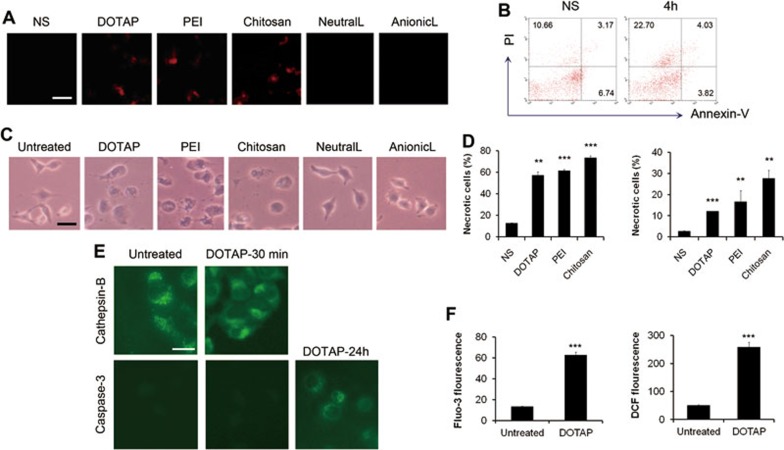
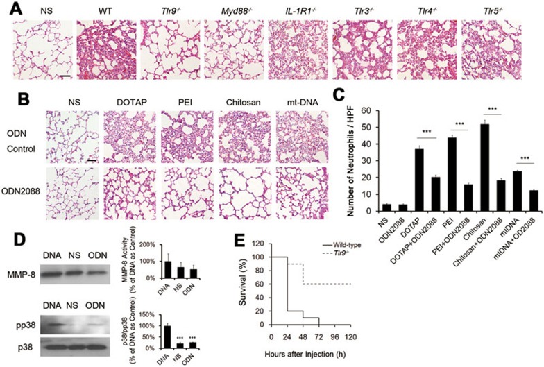
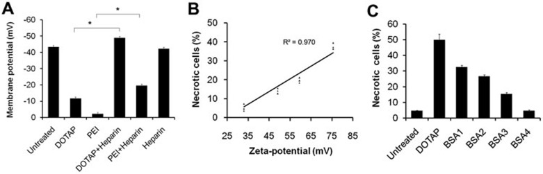
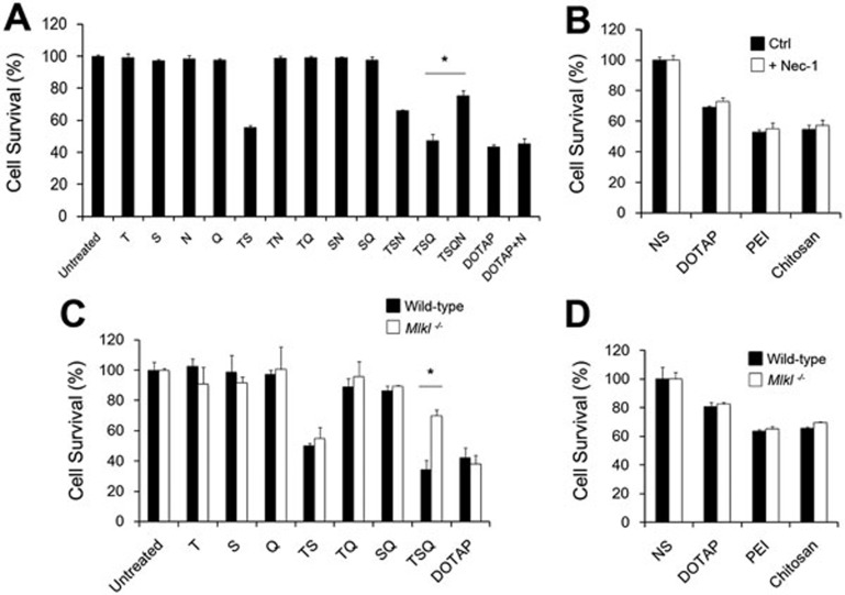
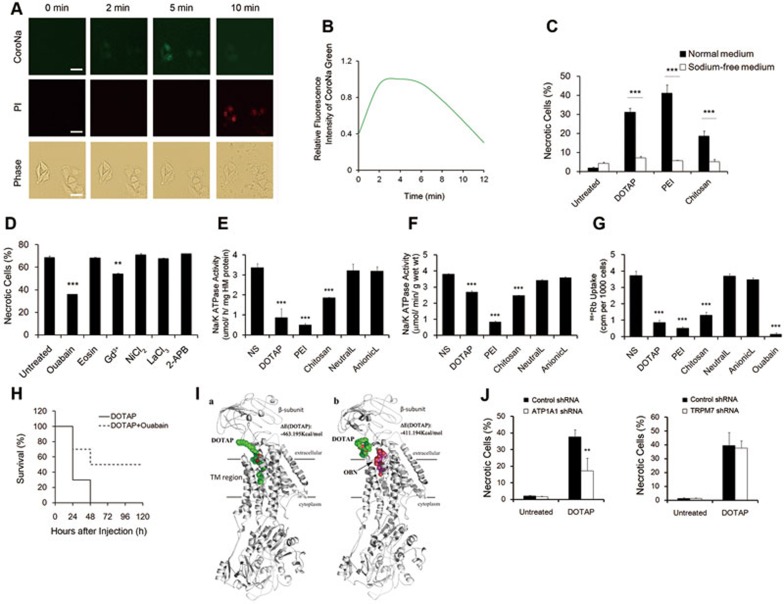
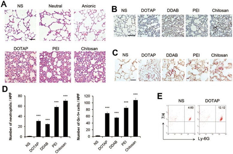
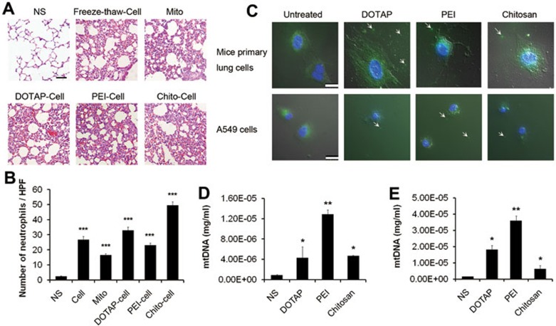
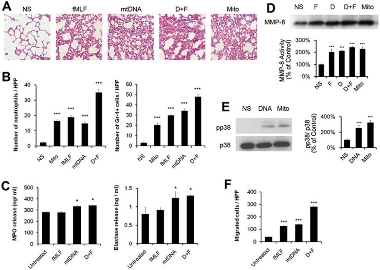
Nanocarriers with positive surface charges are known for their toxicity which has limited their clinical applications. The mechanism underlying their toxicity, such as the induction of inflammatory response, remains largely unknown. In the present study we found that injection of cationic nanocarriers, including cationic liposomes, PEI, and chitosan, led to the rapid appearance of necrotic cells. Cell necrosis induced by cationic nanocarriers is dependent on their positive surface charges, but does not require RIP1 and Mlkl. Instead, intracellular Na(+) overload was found to accompany the cell death. Depletion of Na(+) in culture medium or pretreatment of cells with the Na(+)/K(+)-ATPase cation-binding site inhibitor ouabain, protected cells from cell necrosis. Moreover, treatment with cationic nanocarriers inhibited Na(+)/K(+)-ATPase activity both in vitro and in vivo. The computational simulation showed that cationic carriers could interact with cation-binding site of Na(+)/K(+)-ATPase. Mice pretreated with a small dose of ouabain showed improved survival after injection of a lethal dose of cationic nanocarriers. Further analyses suggest that cell necrosis induced by cationic nanocarriers and the resulting leakage of mitochondrial DNA could trigger severe inflammation in vivo, which is mediated by a pathway involving TLR9 and MyD88 signaling. Taken together, our results reveal a novel mechanism whereby cationic nanocarriers induce acute cell necrosis through the interaction with Na(+)/K(+)-ATPase, with the subsequent exposure of mitochondrial damage-associated molecular patterns as a key event that mediates the inflammatory responses. Our study has important implications for evaluating the biocompatibility of nanocarriers and designing better and safer ones for drug delivery.
表面带正电荷的纳米载体因其毒性而闻名,这限制了它们的临床应用。其毒性背后的机制,如炎症反应的诱导,在很大程度上仍不清楚。在本研究中,我们发现注射阳离子纳米载体,包括阳离子脂质体、聚乙烯亚胺和壳聚糖,会导致坏死细胞迅速出现。阳离子纳米载体诱导的细胞坏死取决于其正表面电荷,但不需要RIP1和Mlkl。相反,发现细胞内钠离子过载伴随着细胞死亡。培养基中钠离子的耗尽或用钠钾ATP酶阳离子结合位点抑制剂哇巴因对细胞进行预处理,可保护细胞免于坏死。此外,阳离子纳米载体处理在体外和体内均抑制钠钾ATP酶活性。计算模拟表明,阳离子载体可与钠钾ATP酶的阳离子结合位点相互作用。用小剂量哇巴因预处理的小鼠在注射致死剂量的阳离子纳米载体后存活率提高。进一步分析表明,阳离子纳米载体诱导的细胞坏死以及由此导致的线粒体DNA泄漏可在体内引发严重炎症,这是由涉及TLR9和MyD88信号传导的途径介导的。综上所述,我们的结果揭示了一种新机制,即阳离子纳米载体通过与钠钾ATP酶相互作用诱导急性细胞坏死,随后线粒体损伤相关分子模式的暴露是介导炎症反应的关键事件。我们的研究对于评估纳米载体的生物相容性以及设计更好、更安全的药物递送纳米载体具有重要意义。