Ryan Julie L, Morgan Douglas R, Dominguez Ricardo L, Thorne Leigh B, Elmore Sandra H, Mino-Kenudson Mari, Lauwers Gregory Y, Booker Jessica K, Gulley Margaret L
Department of Dermatology, University of Rochester Medical Center, Rochester, NY, USA.
Lab Invest. 2009 Jan;89(1):80-90. doi: 10.1038/labinvest.2008.103. Epub 2008 Nov 10.
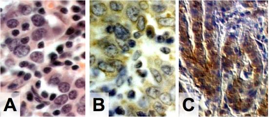
Gastric adenocarcinoma is the second leading cause of cancer death worldwide. Epstein-Barr virus (EBV) is present in the malignant cells of approximately 10% of cases. It is unclear whether EBV is being missed in some gastric adenocarcinomas due to insensitive test methods or partial EBV genome loss. In this study, we screened 113 gastric adenocarcinomas from low- and high-incidence regions (United States and Central America) for the presence of EBV using a battery quantitative real-time PCR (Q-PCR) assays targeting disparate segments of the EBV genome (BamH1W, EBNA1, LMP1, LMP2, BZLF1, EBER1) and histochemical stains targeting EBV-encoded RNA (EBER), the latent proteins LMP1 and LMP2, and the lytic proteins BMRF1 and BZLF1. EBV DNA was detected by Q-PCR in 48/75 United States cancers (64%) and in 38/38 Central American cancers (100%), which was a significant difference. EBER was localized to malignant epithelial cells in 8/48 (17%) United States and 3/38 (8%) Central American cancers. Viral loads were considerably higher for EBER-positive vs EBER-negative cancers (mean 162 986 vs 62 EBV DNA copies per 100,000 cells). A viral load of 2000 copies per 100,000 cells is recommended as the threshold distinguishing EBER-positive from EBER-negative tumors. One infected cancer selectively failed to amplify the LMP2 gene because of a point mutation, whereas another cancer had an atypical pattern of Q-PCR positivity suggesting deletion of large segments of the EBV genome. Three different viral latency profiles were observed in the cancers based on constant expression of EBER and focal or variable expression of LMP1 or LMP2, without lytic protein expression. We conclude that EBV DNA levels generally reflect EBER status, and a panel of at least two Q-PCR assays is recommended for sensitive identification of infected cancers.
胃腺癌是全球癌症死亡的第二大主要原因。爱泼斯坦-巴尔病毒(EBV)存在于约10%的病例的恶性细胞中。目前尚不清楚,是由于检测方法不敏感还是部分EBV基因组缺失,导致某些胃腺癌中EBV被漏检。在本研究中,我们使用一组针对EBV基因组不同片段(BamH1W、EBNA1、LMP1、LMP2、BZLF1、EBER1)的定量实时PCR(Q-PCR)检测方法,以及针对EBV编码RNA(EBER)、潜伏蛋白LMP1和LMP2、裂解蛋白BMRF1和BZLF1的组织化学染色,对来自低发和高发地区(美国和中美洲)的113例胃腺癌进行了EBV检测。通过Q-PCR在美国的75例癌症中检测到48例(64%)EBV DNA,在中美洲的38例癌症中检测到38例(100%),差异显著。在美国的48例癌症中有8例(17%)、中美洲的38例癌症中有3例(8%)检测到EBER定位于恶性上皮细胞。EBER阳性癌症的病毒载量明显高于EBER阴性癌症(平均每100,000个细胞中分别为162 986和62个EBV DNA拷贝)。建议将每100,000个细胞中2000个拷贝的病毒载量作为区分EBER阳性和EBER阴性肿瘤的阈值。1例受感染的癌症因点突变而选择性地未能扩增LMP2基因,而另1例癌症具有非典型的Q-PCR阳性模式,提示EBV基因组大片段缺失。基于EBER的持续表达以及LMP1或LMP2的局灶性或可变表达,且无裂解蛋白表达,在癌症中观察到三种不同的病毒潜伏模式。我们得出结论,EBV DNA水平通常反映EBER状态,建议至少使用两种Q-PCR检测方法进行组合,以灵敏地鉴定受感染的癌症。