Nuclear Medicine Unit,1 Imaging Department, Centre Hospitalier Lyon-Sud, Pierre-Bénite, France.
J Appl Clin Med Phys. 2012 Sep 6;13(5):3875. doi: 10.1120/jacmp.v13i5.3875.
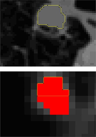
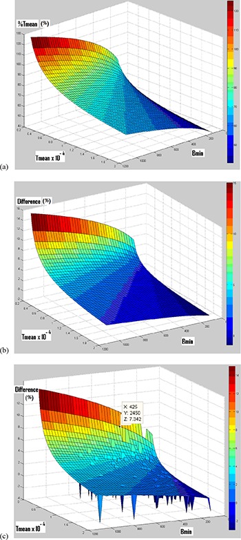
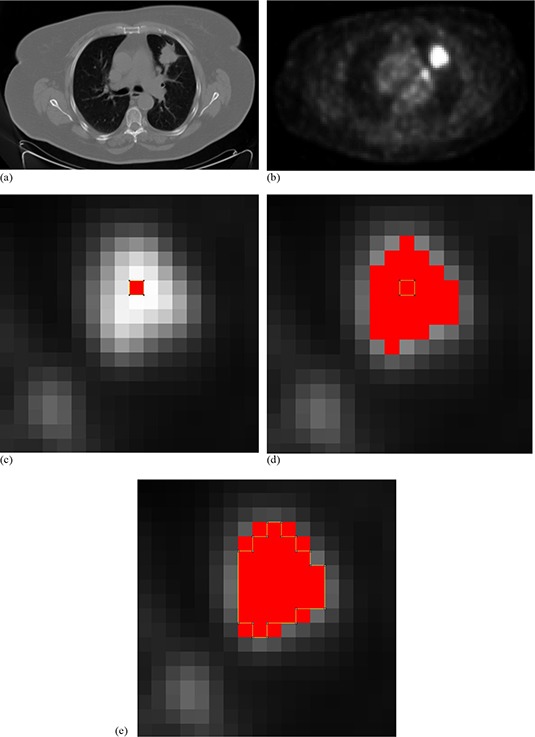
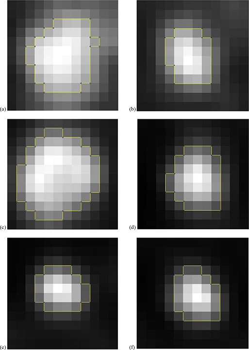
Tumor delineation is a critical aspect in radiotherapy treatment planning and is usually performed with the anatomical images of a computed tomography (CT) scan. For non-small cell lung cancer, it has been recommended to use functional positron emission tomography (PET) images to take into account the biological target characteristics. However, today, there is no satisfactory segmentation technique for PET images in clinical applications. In the present study, a solution to this problem is proposed. The development of the segmentation technique is based on the threshold's adjustment directly from patients, rather than from phantoms. To this end, two references were chosen: measurements performed on CT images of the selected lesions, and histological measurements of surgically removed tumors. The inclusion and exclusion criteria were chosen to produce references that are assumed to have measured tumor sizes equal to the true in vivo tumor sizes. In total, for the two references, 65 lung lesions of 54 patients referred for FDG-PET/CT exams were selected. For validation, measurements of segmented lesions on PET images using this technique were also compared to CT and histological measurements. For lesions greater than 20 mm, our segmentation technique showed a good estimation of histological measurements (mean difference between measured and calculated data equal to -0.8 ± 9.0%) and an acceptable estimation of CT measurements. For lesions smaller than or equal to 20 mm, the method showed disagreement with the measurements derived from histological or CT data. This novel segmentation technique shows high accuracy for the lesions with largest axes between 2 and 4.5 cm. However, it does not correctly evaluate smaller lesions, likely due to the partial volume effect and/or respiratory motions.
肿瘤勾画是放射治疗计划中的一个关键环节,通常使用计算机断层扫描(CT)的解剖图像进行。对于非小细胞肺癌,建议使用功能正电子发射断层扫描(PET)图像来考虑生物靶区的特征。然而,目前在临床应用中,PET 图像还没有令人满意的分割技术。在本研究中,提出了一种解决该问题的方法。该分割技术的开发是基于直接从患者调整阈值,而不是从体模进行。为此,选择了两个参考标准:对选定病变的 CT 图像进行测量,以及对手术切除的肿瘤进行组织学测量。选择纳入和排除标准是为了产生被认为测量的肿瘤大小与真实体内肿瘤大小相等的参考标准。总共为这两个参考标准,选择了 54 名接受 FDG-PET/CT 检查的患者的 65 个肺部病变。为了验证,还将使用该技术对 PET 图像上分割病变的测量值与 CT 和组织学测量值进行比较。对于大于 20mm 的病变,我们的分割技术对组织学测量值的估计较好(测量值与计算值之间的平均差异等于-0.8±9.0%),对 CT 测量值的估计也可以接受。对于小于或等于 20mm 的病变,该方法与从组织学或 CT 数据得出的测量值存在分歧。对于最长轴在 2 至 4.5cm 之间的病变,这种新的分割技术具有较高的准确性。然而,它不能正确评估较小的病变,可能是由于部分容积效应和/或呼吸运动的影响。