Guberman Jonathan M, Fay Allison, Dworkin Jonathan, Wingreen Ned S, Gitai Zemer
Department of Molecular Biology, Princeton University, Princeton, New Jersey, United States of America.
PLoS Comput Biol. 2008 Nov;4(11):e1000233. doi: 10.1371/journal.pcbi.1000233. Epub 2008 Nov 28.
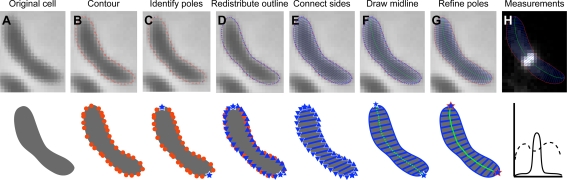
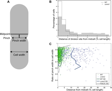
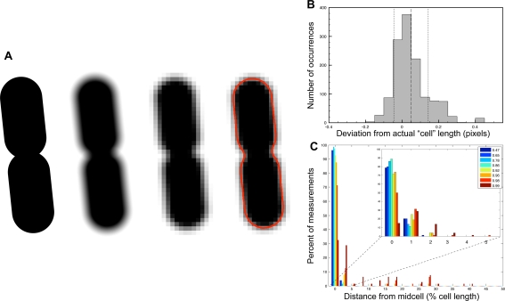
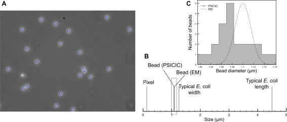
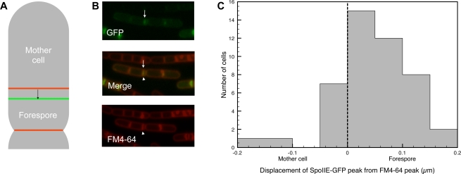
Live-cell imaging by light microscopy has demonstrated that all cells are spatially and temporally organized. Quantitative, computational image analysis is an important part of cellular imaging, providing both enriched information about individual cell properties and the ability to analyze large datasets. However, such studies are often limited by the small size and variable shape of objects of interest. Here, we address two outstanding problems in bacterial cell division by developing a generally applicable, standardized, and modular software suite termed Projected System of Internal Coordinates from Interpolated Contours (PSICIC) that solves common problems in image quantitation. PSICIC implements interpolated-contour analysis for accurate and precise determination of cell borders and automatically generates internal coordinate systems that are superimposable regardless of cell geometry. We have used PSICIC to establish that the cell-fate determinant, SpoIIE, is asymmetrically localized during Bacillus subtilis sporulation, thereby demonstrating the ability of PSICIC to discern protein localization features at sub-pixel scales. We also used PSICIC to examine the accuracy of cell division in Esherichia coli and found a new role for the Min system in regulating division-site placement throughout the cell length, but only prior to the initiation of cell constriction. These results extend our understanding of the regulation of both asymmetry and accuracy in bacterial division while demonstrating the general applicability of PSICIC as a computational approach for quantitative, high-throughput analysis of cellular images.
通过光学显微镜进行的活细胞成像表明,所有细胞在空间和时间上都是有组织的。定量计算图像分析是细胞成像的重要组成部分,它既能提供有关单个细胞特性的丰富信息,又具备分析大型数据集的能力。然而,此类研究常常受到感兴趣对象尺寸小和形状多变的限制。在此,我们通过开发一套通用、标准化且模块化的软件套件——插值轮廓内部坐标投影系统(PSICIC),解决图像定量中的常见问题,从而应对细菌细胞分裂中的两个突出问题。PSICIC实现了插值轮廓分析,用于准确精确地确定细胞边界,并自动生成无论细胞几何形状如何都可叠加的内部坐标系。我们利用PSICIC确定了细胞命运决定因子SpoIIE在枯草芽孢杆菌孢子形成过程中不对称定位,从而证明了PSICIC在亚像素尺度上辨别蛋白质定位特征的能力。我们还使用PSICIC检查了大肠杆菌细胞分裂的准确性,发现Min系统在调节整个细胞长度上的分裂位点定位方面有新作用,但仅在细胞缢缩开始之前。这些结果扩展了我们对细菌分裂中不对称性和准确性调控的理解,同时证明了PSICIC作为一种用于细胞图像定量高通量分析的计算方法具有普遍适用性。