Adult Congenital Heart Programme, Department of Cardiology, Royal Brompton & Harefield NHS Trust, Imperial College, London, UK.
Int J Cardiol. 2010 Mar 18;139(3):283-8. doi: 10.1016/j.ijcard.2008.10.043. Epub 2008 Dec 6.
Repaired coarctation of the aorta is associated with premature atherosclerosis and an increased risk of cardiovascular events even in normotensive subjects. To date clinical risk stratification has focused on brachial blood pressures ignoring the complex pulsatility of the aortic wave form. The aim of this study was to assess components of this pulsatility in a clinical setting and to suggest possible techniques to improve risk stratification.
This was a prospective study recruiting patients from a tertiary referral centre. Pulse wave morphology was assessed using applanation tonometry. B-mode ultrasound and cardiac magnetic resonance were used to assess carotid intimal-medial thickness and left ventricular mass.
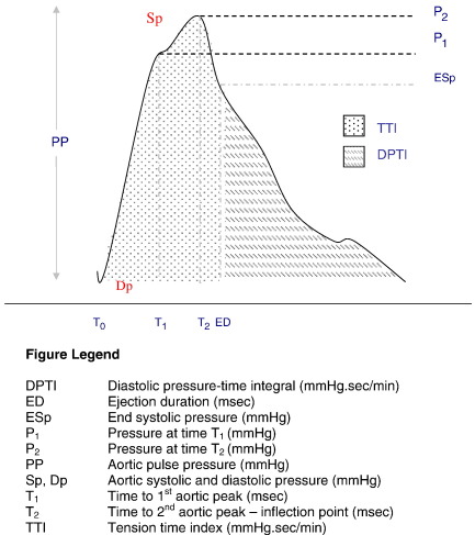
Forty-six subjects with repaired coarctation of the aorta (range 16-62 years; mean 31 years) and 20 matched controls were studied. Baseline brachial systolic and diastolic blood pressures were not statistically different between the 2 groups. Peripheral (62.5 mmHg (11.3) vs. 50.6 mmHg (15.0), p=0.0008) and central (34.5 mmHg (7.7) vs. 28.7 mmHg (4.7), p=0.005) pulse pressures were elevated in the coarctation patients compared to controls. The reflected pressure wave returned to the ascending aorta earlier in the coarctation group (p=0.007) and the tension time index (TTI) was increased (p=0.03). The sub-endocardial viability index (SVI) was reduced in the coarctation subjects (159 (33) vs. 186 (31)%; p=0.009) but there was no differences in central augmentation index (p=0.35).
This study demonstrates that there are patients with repaired coarctation who have an excellent mid-term outcome free from ventricular hypertrophy, carotid intima medial thickening and with relatively preserved vascular reactivity. However even in this "best outcome" cohort there were abnormal vascular characteristics that may predispose to increased cardiovascular risk. Simple non-invasive investigations can more extensively characterise these sub-clinical abnormalities and by utilised in long-term surveillance.
即使在血压正常的患者中,修复后的主动脉缩窄也与过早的动脉粥样硬化和心血管事件风险增加相关。迄今为止,临床风险分层主要集中在肱动脉血压上,而忽略了主动脉波形态的复杂搏动性。本研究旨在评估这种搏动性在临床环境中的组成部分,并提出可能改善风险分层的技术。
这是一项前瞻性研究,从三级转诊中心招募患者。使用平板张力计评估脉搏波形态。B 型超声和心脏磁共振用于评估颈动脉内膜中层厚度和左心室质量。
研究共纳入 46 例修复后的主动脉缩窄患者(年龄 16-62 岁,平均 31 岁)和 20 名匹配的对照者。两组患者的基线肱动脉收缩压和舒张压无统计学差异。与对照组相比,外周(62.5mmHg(11.3)vs.50.6mmHg(15.0),p=0.0008)和中心(34.5mmHg(7.7)vs.28.7mmHg(4.7),p=0.005)脉压升高。反射波更早地回到升主动脉在缩窄组(p=0.007),张力时间指数(TTI)增加(p=0.03)。在缩窄患者中,心内膜下活力指数(SVI)降低(159(33)vs.186(31)%,p=0.009),但中心增强指数无差异(p=0.35)。
本研究表明,有一些修复后的主动脉缩窄患者具有良好的中期预后,没有心室肥厚、颈动脉内膜中层增厚,并且血管反应性相对保留。然而,即使在这种“最佳结局”的队列中,仍存在可能增加心血管风险的异常血管特征。简单的非侵入性检查可以更广泛地描述这些亚临床异常,并在长期监测中使用。