Nair Sandeep P, Shiau Deng-Shan, Principe Jose C, Iasemidis Leonidas D, Pardalos Panos M, Norman Wendy M, Carney Paul R, Kelly Kevin M, Sackellares J Chris
Department of Neurology, Allegheny General Hospital, Center for Neuroscience Research, Allegheny-Singer Research Intitute, Pittsburgh, PA, USA.
Exp Neurol. 2009 Mar;216(1):115-21. doi: 10.1016/j.expneurol.2008.11.009. Epub 2008 Nov 27.
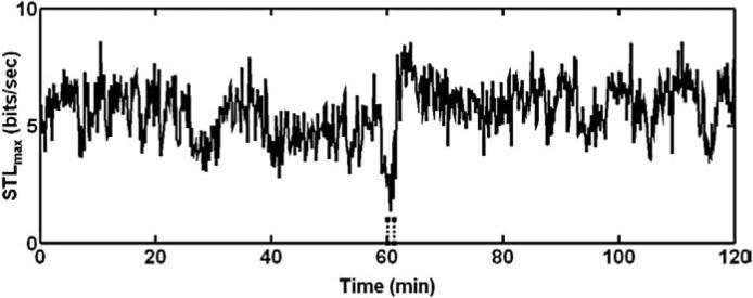
Analysis of intracranial electroencephalographic (iEEG) recordings in patients with temporal lobe epilepsy (TLE) has revealed characteristic dynamical features that distinguish the interictal, ictal, and postictal states and inter-state transitions. Experimental investigations into the mechanisms underlying these observations require the use of an animal model. A rat TLE model was used to test for differences in iEEG dynamics between well-defined states and to test specific hypotheses: 1) the short-term maximum Lyapunov exponent (STL(max)), a measure of signal order, is lowest and closest in value among cortical sites during the ictal state, and highest and most divergent during the postictal state; 2) STL(max) values estimated from the stimulated hippocampus are the lowest among all cortical sites; and 3) the transition from the interictal to ictal state is associated with a convergence in STL(max) values among cortical sites. iEEGs were recorded from bilateral frontal cortices and hippocampi. STL(max) and T-index (a measure of convergence/divergence of STL(max) between recorded brain areas) were compared among the four different periods. Statistical tests (ANOVA and multiple comparisons) revealed that ictal STL(max) was lower (p<0.05) than other periods, STL(max) values corresponding to the stimulated hippocampus were lower than those estimated from other cortical regions, and T-index values were highest during the postictal period and lowest during the ictal period. Also, the T-index values corresponding to the preictal period were lower than those during the interictal period (p<0.05). These results indicate that a rat TLE model demonstrates several important dynamical signal characteristics similar to those found in human TLE and support future use of the model to study epileptic state transitions.
对颞叶癫痫(TLE)患者颅内脑电图(iEEG)记录的分析揭示了区分发作间期、发作期和发作后期状态以及状态间转换的特征性动力学特征。对这些观察结果背后机制的实验研究需要使用动物模型。使用大鼠TLE模型来测试明确状态之间iEEG动力学的差异,并检验特定假设:1)短期最大Lyapunov指数(STL(max)),一种信号秩序的度量,在发作期皮质部位中最低且值最接近,在发作后期最高且差异最大;2)从受刺激海马体估计的STL(max)值在所有皮质部位中是最低的;3)从发作间期到发作期的转变与皮质部位间STL(max)值的收敛有关。从双侧额叶皮质和海马体记录iEEG。在四个不同时期比较STL(max)和T指数(记录脑区之间STL(max)收敛/发散的度量)。统计检验(方差分析和多重比较)显示,发作期STL(max)低于其他时期(p<0.05),对应受刺激海马体的STL(max)值低于从其他皮质区域估计的值,T指数值在发作后期最高,在发作期最低。此外,发作前期对应的T指数值低于发作间期(p<0.05)。这些结果表明,大鼠TLE模型表现出与人类TLE中发现的几个重要动力学信号特征相似,并支持该模型未来用于研究癫痫状态转换。