Noszczyk-Nowak Agnieszka, Nicpoń Józef, Nowak Marcin, Slawuta Piotr
Department of Internal and Parasitic Diseases Veterinary Medicine Faculty, Wroclaw University of Environmental and Life Sciences, Wrocław 50-366, Poland.
Acta Vet Scand. 2009 Jan 30;51(1):6. doi: 10.1186/1751-0147-51-6.
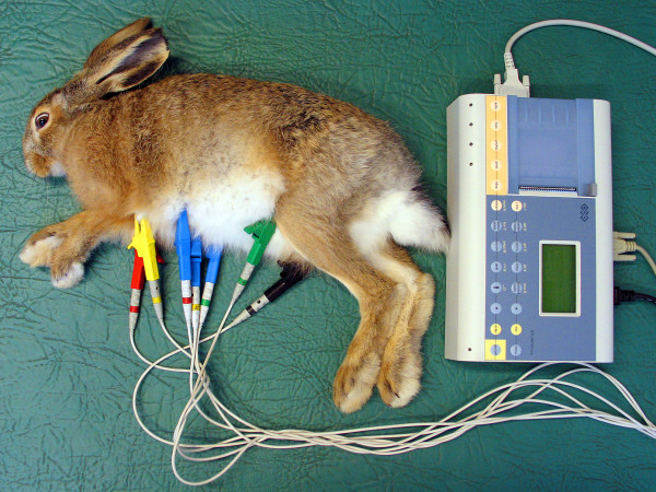
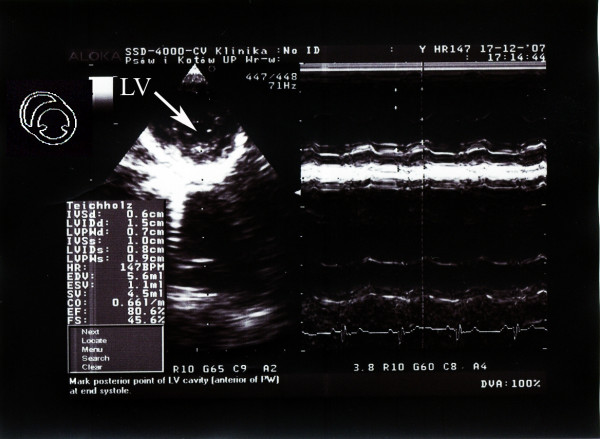
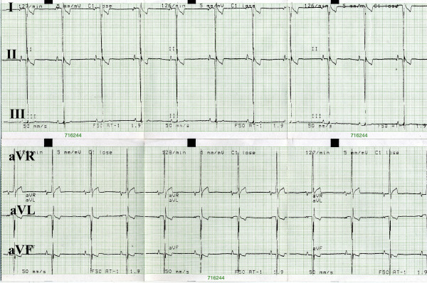
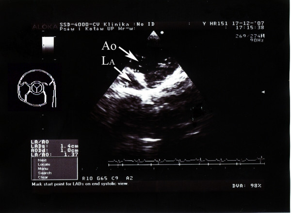
The study aimed at defining reference values for electrocardiographic (ECG) and echocardiographic parameters as well as macroscopic dimensions of the heart and microscopic dimensions of cardiomyocytes in the European brown hare. The studies were conducted on 30 adult, clinically healthy hares of either sex caught in Poland. ECG and echocardiography were performed supravitally on anaesthetized hares. After euthanasia, gross and microscopic myocardial and cardiomyocyte dimensions were determined. Heart rate amounted to 140 +/- 37.5 beats/min, the leading rhythm involved the sinus rhythm. P wave time was 26 +/- 5 ms, PQ time was 80 ms, QRS time was 29 +/- 3.5 ms, and ST was 97.5 +/- 7 ms. Echocardiography determined a left ventricular wall end-diastolic diameter of 8.6 +/- 2.0 mm and an intraventricular septum end-diastolic diameter of 5.75 +/- 1.0 mm. The thickness of the interventricular septum corresponded to that of the free wall of the left ventricle, a finding consistent with physiological hypertrophy. Preliminary reference values were established for echocardiography. The findings were similar to those obtained at necropsy. The ECG and echocardiographic studies represent the first supravital examination of cardiac function in the hare. The obtained results illustrate adaptation of hare's myocardium to its mode of life. The cardiac findings resemble the athlete's heart syndrome described in humans. The findings may prove useful in further studies on the physiology of the cardio-vascular system in the hare.
该研究旨在确定欧洲棕兔心电图(ECG)和超声心动图参数的参考值,以及心脏的宏观尺寸和心肌细胞的微观尺寸。研究对象为在波兰捕获的30只成年、临床健康的雌雄野兔。在麻醉的野兔身上进行活体心电图和超声心动图检查。安乐死后,测定心脏大体和微观的心肌及心肌细胞尺寸。心率为140±37.5次/分钟,主导节律为窦性节律。P波时间为26±5毫秒,PQ时间为80毫秒,QRS时间为29±3.5毫秒,ST段为97.5±7毫秒。超声心动图测定左心室壁舒张末期直径为8.6±2.0毫米,室间隔舒张末期直径为5.75±1.0毫米。室间隔厚度与左心室游离壁厚度相当,这一发现与生理性肥厚相符。建立了超声心动图的初步参考值。这些结果与尸检结果相似。心电图和超声心动图研究是首次对野兔心脏功能进行的活体检查。所获结果说明了野兔心肌对其生活方式的适应性。心脏检查结果类似于人类描述的运动员心脏综合征。这些发现可能对进一步研究野兔心血管系统生理学有用。