Freeburg Elizabeth M, Goyeneche Alicia A, Telleria Carlos M
Sanford School of Medicine, The University of South Dakota, Vermillion, SD 57069, USA.
Int J Oncol. 2009 Mar;34(3):743-55. doi: 10.3892/ijo_00000200.
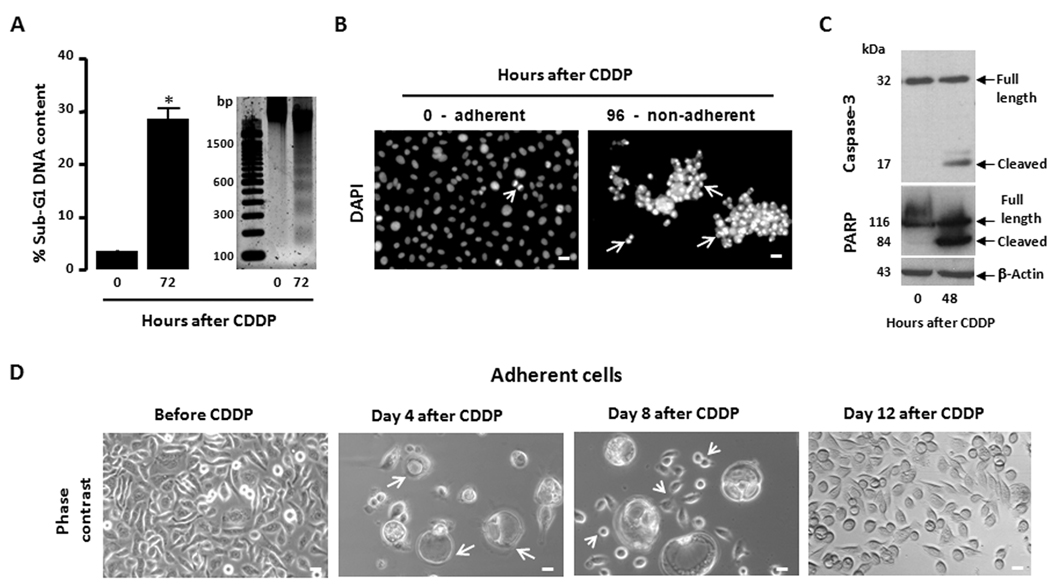
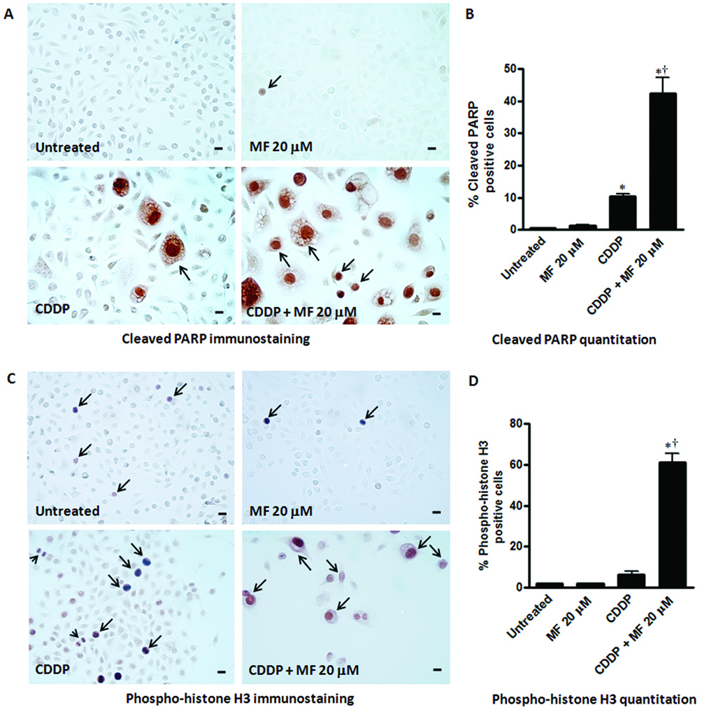
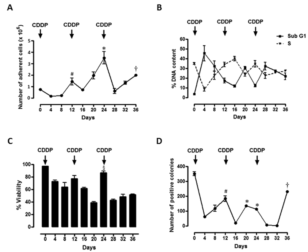
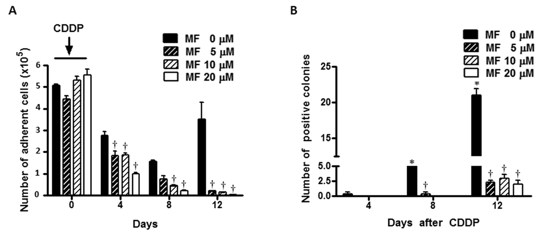
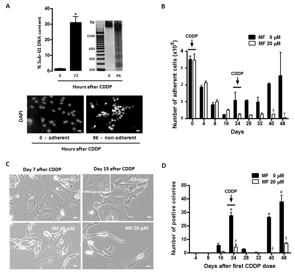
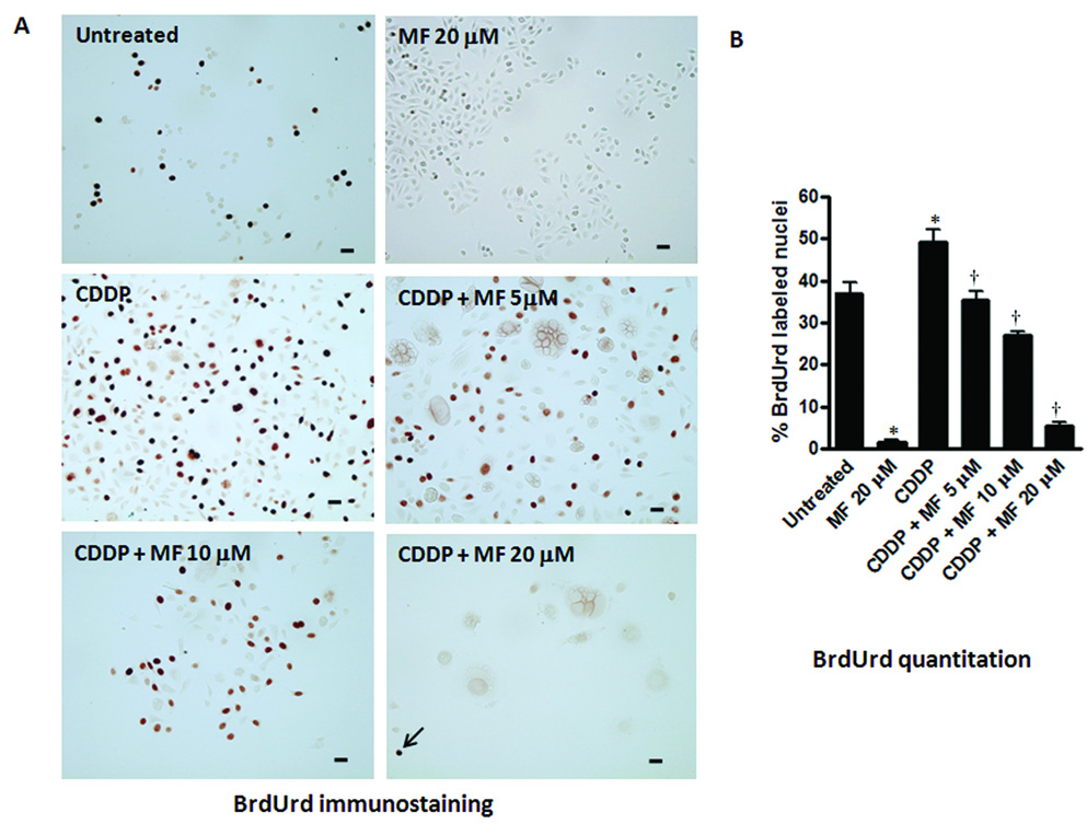
Repopulation of cancer cells escaping lethal chemotherapy is a critical factor hindering treatment success. One strategy to inhibit tumor cell repopulation is the use of cytostatic compounds between courses of lethal chemotherapy. In this study, we tested the hypothesis that mifepristone (MF), a steroid compound with demonstrated growth inhibition activity in ovarian cancer, should be efficacious in inducing cytostasis and preventing repopulation of ovarian cancer cells if given among rounds of cisplatin (CDDP) treatment. We established an in vitro approach wherein ovarian cancer cells with high (OV2008) or low (SK-OV-3) sensitivity to CDDP were exposed to 3 (OV2008) or 2 (SK-OV-3) rounds of lethal doses of CDDP for 1 h, 12 (OV2008) or 24 (SK-OV-3) days apart. Every 4 or 8 days cell number, cell viability, cell cycle traverse, and colony-forming capacity of viable cells was analyzed. Although CDDP killed the vast majority of cells, there were remnant cells escaping CDDP lethality and repopulating the culture, as evidenced by increased cell number, improved clonogenic capacity of viable cells, and normalization of DNA synthesis. Conversely, when cells were exposed to CDDP for 1 h, and 5, 10 or 20 microM MF was present in the culture medium after CDDP removal, the number, clonogenic capacity, and DNA synthesis ability of the cells were reduced in a dose-dependent manner. The blockage by MF of post-CDDP repopulation was accompanied by a remarkable increase in the percentage of cells expressing the cell death marker cleaved poly(ADP-ribose) polymerase and the mitotic marker phospho-histone H3, suggesting that MF also potentiated CDDP lethality and that the cells likely die due to mitotic failure. In summary, this is the first study reporting that presence of cytostatic concentrations of MF after courses of lethal doses of CDDP prevents repopulation of remnant ovarian cancer cells surviving CDDP treatment.
逃避致死性化疗的癌细胞再增殖是阻碍治疗成功的关键因素。抑制肿瘤细胞再增殖的一种策略是在致死性化疗疗程之间使用细胞生长抑制剂。在本研究中,我们检验了以下假设:米非司酮(MF)是一种在卵巢癌中具有已证实的生长抑制活性的类固醇化合物,如果在顺铂(CDDP)治疗轮次之间给予,应能有效诱导细胞生长停滞并防止卵巢癌细胞再增殖。我们建立了一种体外方法,将对CDDP高敏(OV2008)或低敏(SK-OV-3)的卵巢癌细胞分别暴露于3(OV2008)或2(SK-OV-3)轮致死剂量的CDDP中1小时,间隔12(OV2008)或24(SK-OV-3)天。每4或8天分析活细胞的细胞数量、细胞活力、细胞周期进程和集落形成能力。尽管CDDP杀死了绝大多数细胞,但仍有残余细胞逃避CDDP致死性并使培养物再增殖,这表现为细胞数量增加、活细胞克隆形成能力提高以及DNA合成正常化。相反,当细胞暴露于CDDP 1小时后,在去除CDDP后培养基中存在5、10或20微摩尔的MF时,细胞的数量、克隆形成能力和DNA合成能力以剂量依赖方式降低。MF对CDDP后再增殖的阻断伴随着表达细胞死亡标志物裂解的聚(ADP-核糖)聚合酶和有丝分裂标志物磷酸化组蛋白H3的细胞百分比显著增加,这表明MF也增强了CDDP的致死性,并且细胞可能因有丝分裂失败而死亡。总之,这是第一项报道在致死剂量的CDDP疗程后存在细胞生长抑制浓度的MF可防止CDDP治疗后存活的残余卵巢癌细胞再增殖的研究。