Walsh Naomi, O'Donovan Norma, Kennedy Susan, Henry Michael, Meleady Paula, Clynes Martin, Dowling Paul
National Institute for Cellular Biotechnology, Dublin City University, Glasnevin, Ireland, UK.
Proteome Sci. 2009 Feb 14;7:3. doi: 10.1186/1477-5956-7-3.
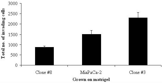
Markers of pancreatic cancer invasion were investigated in two clonal populations of the cell line, MiaPaCa-2, Clone #3 (high invasion) and Clone #8 (low invasion) using proteomic profiling of an in vitro model of pancreatic cancer.
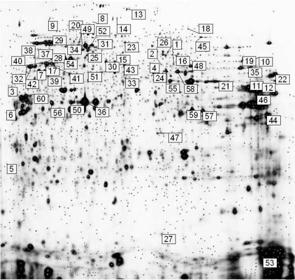
Using 2D-DIGE followed by MALDI-TOF MS, two clonal sub-populations of the pancreatic cancer cell line, MiaPaCa-2 with high and low invasive capacities were incubated on matrigel 24 hours prior to analysis to stimulate cell-ECM contact and mimic in vivo interaction with the basement membrane.
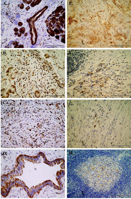
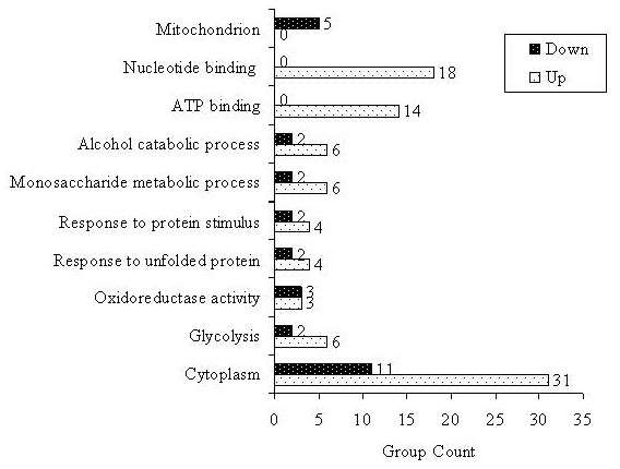
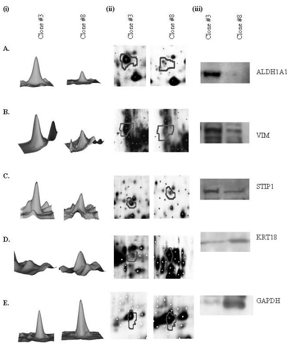
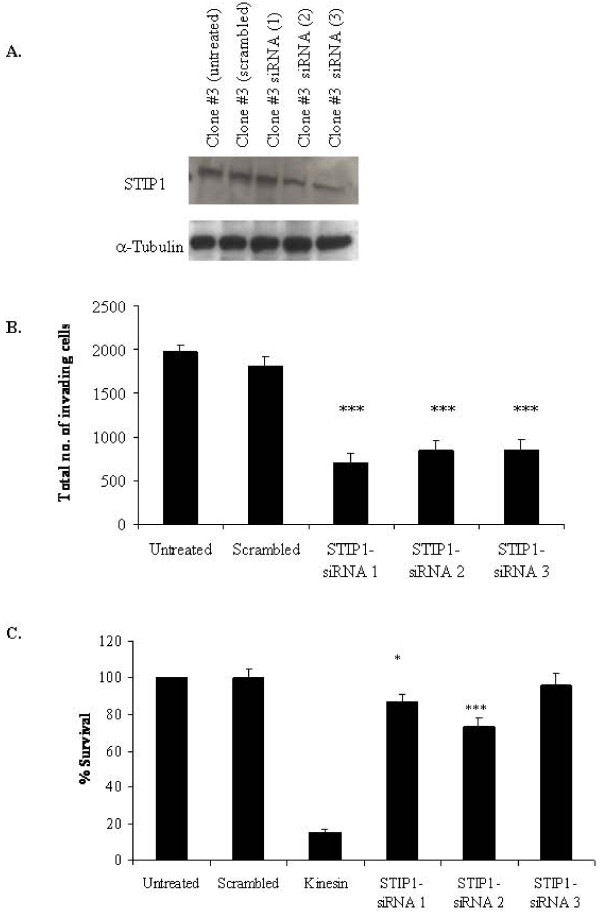
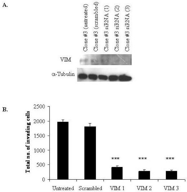
Sixty proteins were identified as being differentially expressed (> 1.2 fold change and p < or = 0.05) between Clone #3 and Clone #8. Proteins found to have higher abundance levels in the highly invasive Clone #3 compared to the low invasive Clone #8 include members of the chaperone activity proteins and cytoskeleton constituents whereas metabolism-associated and catalytic proteins had lower abundance levels. Differential protein expression levels of ALDH1A1, VIM, STIP1 and KRT18 and GAPDH were confirmed by immunoblot. Using RNAi technology, STIP1 knockdown significantly reduced invasion and proliferation of the highly invasive Clone #3. Knockdown of another target, VIM by siRNA in Clone #3 cells also resulted in decreased invasion abilities of Clone #3. Elevated expression of STIP1 was observed in pancreatic tumour tissue compared to normal pancreas, whereas ALDH1A1 stained at lower levels in pancreatic tumours, as detected by immunohistochemistry.
Identification of targets which play a role in the highly invasive phenotype of pancreatic cancer may help to understand the biological behaviour, the rapid progression of this cancer and may be of importance in the development of new therapeutic strategies for pancreatic cancer.
利用胰腺癌体外模型的蛋白质组学分析,在细胞系MiaPaCa-2的两个克隆群体(克隆#3,高侵袭性;克隆#8,低侵袭性)中研究了胰腺癌侵袭的标志物。
采用二维差异凝胶电泳(2D-DIGE)结合基质辅助激光解吸电离飞行时间质谱(MALDI-TOF MS),将具有高侵袭能力和低侵袭能力的胰腺癌细胞系MiaPaCa-2的两个克隆亚群在基质胶上孵育24小时,然后进行分析,以刺激细胞与细胞外基质(ECM)接触并模拟其在体内与基底膜的相互作用。
在克隆#3和克隆#8之间,鉴定出60种差异表达的蛋白质(变化倍数>1.2且p≤0.05)。与低侵袭性的克隆#8相比,在高侵袭性的克隆#3中丰度水平较高的蛋白质包括伴侣活性蛋白成员和细胞骨架成分,而与代谢相关的蛋白质和催化蛋白的丰度水平较低。通过免疫印迹法证实了醛脱氢酶1A1(ALDH1A1)、波形蛋白(VIM)、应激诱导磷酸化蛋白1(STIP1)、细胞角蛋白18(KRT18)和甘油醛-3-磷酸脱氢酶(GAPDH)的差异蛋白表达水平。利用RNA干扰(RNAi)技术,敲低STIP1可显著降低高侵袭性克隆#3的侵袭和增殖能力。在克隆#3细胞中,通过小干扰RNA(siRNA)敲低另一个靶点VIM,也导致克隆#3的侵袭能力下降。免疫组织化学检测显示,与正常胰腺相比,胰腺癌组织中STIP1表达升高,而ALDH1A1染色水平较低。
鉴定在胰腺癌高侵袭表型中起作用的靶点,可能有助于理解这种癌症的生物学行为、快速进展情况,并且在开发胰腺癌新治疗策略方面可能具有重要意义。