• 文献检索
  • 文档翻译
  • 深度研究
  • 学术资讯
  • Suppr Zotero 插件Zotero 插件
  • 邀请有礼
  • 套餐&价格
  • 历史记录
应用&插件
Suppr Zotero 插件Zotero 插件浏览器插件Mac 客户端Windows 客户端微信小程序
定价
高级版会员购买积分包购买API积分包
服务
文献检索文档翻译深度研究API 文档MCP 服务
关于我们
关于 Suppr公司介绍联系我们用户协议隐私条款
关注我们

Suppr 超能文献

核心技术专利:CN118964589B侵权必究
粤ICP备2023148730 号-1Suppr @ 2026

文献检索

告别复杂PubMed语法,用中文像聊天一样搜索,搜遍4000万医学文献。AI智能推荐,让科研检索更轻松。

立即免费搜索

文件翻译

保留排版,准确专业,支持PDF/Word/PPT等文件格式,支持 12+语言互译。

免费翻译文档

深度研究

AI帮你快速写综述,25分钟生成高质量综述,智能提取关键信息,辅助科研写作。

立即免费体验

基于磁共振成像(MRI)的人类大脑皮质下、脑室及颅内脑容量测量:扫描时段、采集序列、数据分析、扫描仪升级、扫描仪供应商及场强的可靠性影响

MRI-derived measurements of human subcortical, ventricular and intracranial brain volumes: Reliability effects of scan sessions, acquisition sequences, data analyses, scanner upgrade, scanner vendors and field strengths.

作者信息

Jovicich Jorge, Czanner Silvester, Han Xiao, Salat David, van der Kouwe Andre, Quinn Brian, Pacheco Jenni, Albert Marilyn, Killiany Ronald, Blacker Deborah, Maguire Paul, Rosas Diana, Makris Nikos, Gollub Randy, Dale Anders, Dickerson Bradford C, Fischl Bruce

机构信息

Center for Mind-Brain Sciences, Department of Cognitive and Education Sciences, University of Trento, Italy.

出版信息

Neuroimage. 2009 May 15;46(1):177-92. doi: 10.1016/j.neuroimage.2009.02.010. Epub 2009 Feb 20.

DOI:10.1016/j.neuroimage.2009.02.010
PMID:19233293
原文链接:https://pmc.ncbi.nlm.nih.gov/articles/PMC2866077/
Abstract

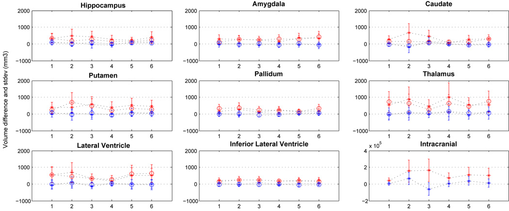
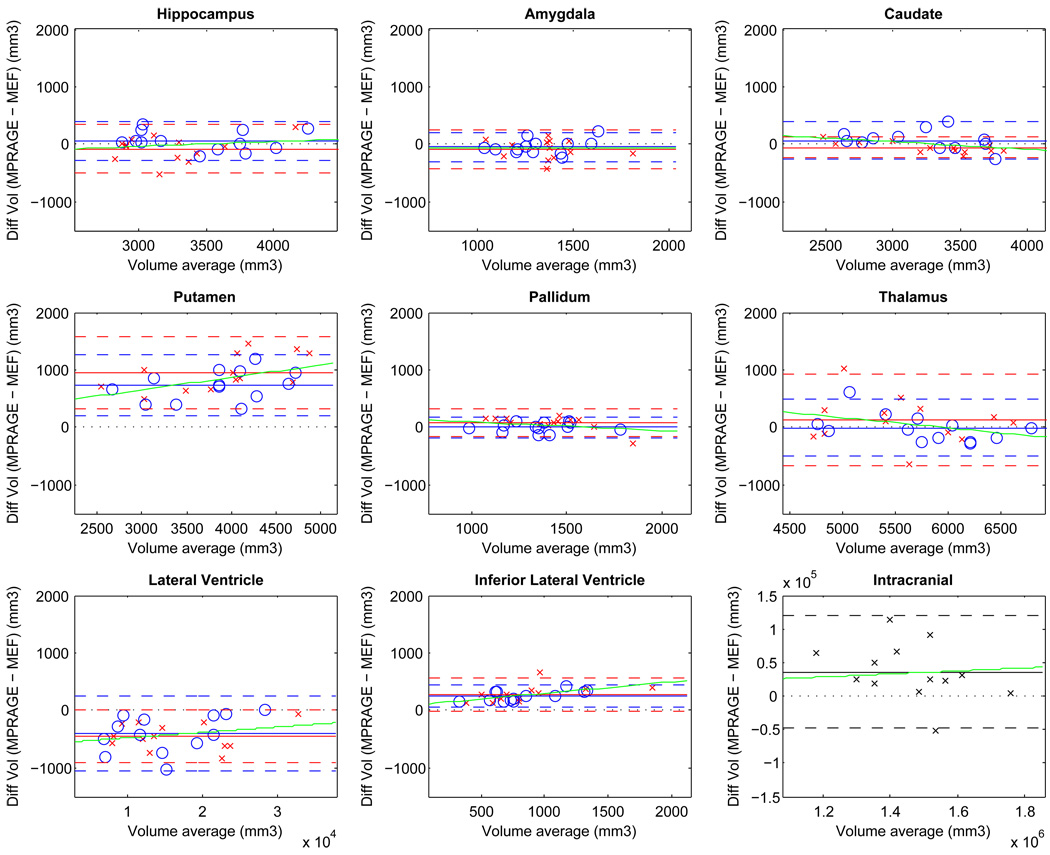
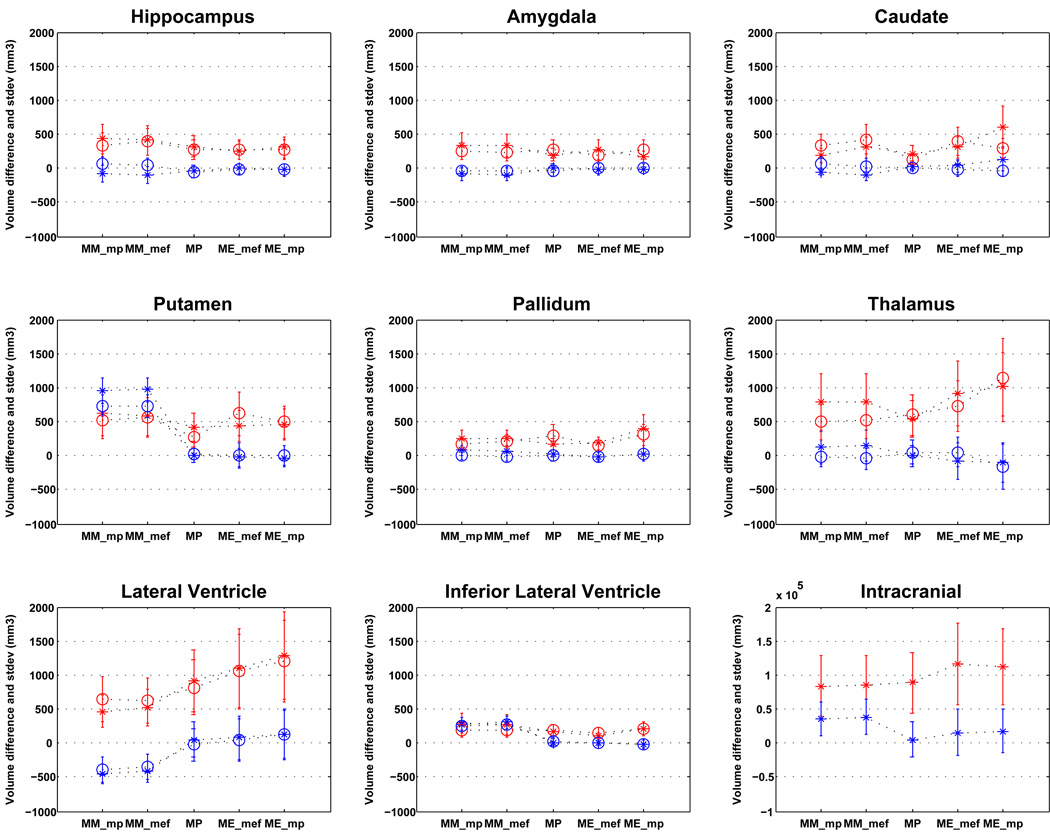
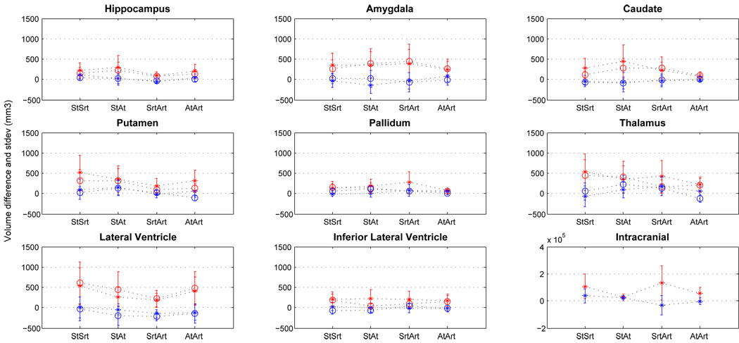
Automated MRI-derived measurements of in-vivo human brain volumes provide novel insights into normal and abnormal neuroanatomy, but little is known about measurement reliability. Here we assess the impact of image acquisition variables (scan session, MRI sequence, scanner upgrade, vendor and field strengths), FreeSurfer segmentation pre-processing variables (image averaging, B1 field inhomogeneity correction) and segmentation analysis variables (probabilistic atlas) on resultant image segmentation volumes from older (n=15, mean age 69.5) and younger (both n=5, mean ages 34 and 36.5) healthy subjects. The variability between hippocampal, thalamic, caudate, putamen, lateral ventricular and total intracranial volume measures across sessions on the same scanner on different days is less than 4.3% for the older group and less than 2.3% for the younger group. Within-scanner measurements are remarkably reliable across scan sessions, being minimally affected by averaging of multiple acquisitions, B1 correction, acquisition sequence (MPRAGE vs. multi-echo-FLASH), major scanner upgrades (Sonata-Avanto, Trio-TrioTIM), and segmentation atlas (MPRAGE or multi-echo-FLASH). Volume measurements across platforms (Siemens Sonata vs. GE Signa) and field strengths (1.5 T vs. 3 T) result in a volume difference bias but with a comparable variance as that measured within-scanner, implying that multi-site studies may not necessarily require a much larger sample to detect a specific effect. These results suggest that volumes derived from automated segmentation of T1-weighted structural images are reliable measures within the same scanner platform, even after upgrades; however, combining data across platform and across field-strength introduces a bias that should be considered in the design of multi-site studies, such as clinical drug trials. The results derived from the young groups (scanner upgrade effects and B1 inhomogeneity correction effects) should be considered as preliminary and in need for further validation with a larger dataset.

摘要

通过自动化磁共振成像(MRI)测量活体人类脑容量,能为正常和异常神经解剖学提供新的见解,但对于测量可靠性却知之甚少。在此,我们评估了图像采集变量(扫描时段、MRI序列、扫描仪升级、供应商和场强)、FreeSurfer分割预处理变量(图像平均、B1场不均匀性校正)以及分割分析变量(概率图谱)对年龄较大(n = 15,平均年龄69.5岁)和年龄较小(均为n = 5,平均年龄分别为34岁和36.5岁)的健康受试者所得图像分割体积的影响。在同一台扫描仪上不同日期进行的各次扫描中,老年组海马体、丘脑、尾状核、壳核、侧脑室和总颅内体积测量值之间的变异性小于4.3%,而年轻组则小于2.3%。在同一台扫描仪内,各次扫描的测量结果非常可靠,受多次采集平均、B1校正、采集序列(MPRAGE与多回波FLASH)、主要扫描仪升级(索纳塔 - 阿凡托、 Trio - TrioTIM)以及分割图谱(MPRAGE或多回波FLASH)的影响极小。跨平台(西门子索纳塔与通用电气Signa)和场强(1.5 T与3 T)的体积测量会导致体积差异偏差,但方差与同一台扫描仪内测量的方差相当,这意味着多中心研究不一定需要大得多的样本量就能检测到特定效应。这些结果表明,即使经过升级,在同一扫描仪平台内,从T1加权结构图像的自动分割得出的体积是可靠的测量值;然而,跨平台和跨场强合并数据会引入偏差,在多中心研究(如临床药物试验)设计中应予以考虑。来自年轻组的结果(扫描仪升级效应和B1不均匀性校正效应)应视为初步结果,需要用更大的数据集进行进一步验证。

https://cdn.ncbi.nlm.nih.gov/pmc/blobs/17ae/2866077/94e923e03058/nihms195969f9.jpg
https://cdn.ncbi.nlm.nih.gov/pmc/blobs/17ae/2866077/0a33d026d3b8/nihms195969f1.jpg
https://cdn.ncbi.nlm.nih.gov/pmc/blobs/17ae/2866077/604da166f56f/nihms195969f2.jpg
https://cdn.ncbi.nlm.nih.gov/pmc/blobs/17ae/2866077/d9040e917097/nihms195969f3.jpg
https://cdn.ncbi.nlm.nih.gov/pmc/blobs/17ae/2866077/8fe951ef693c/nihms195969f4.jpg
https://cdn.ncbi.nlm.nih.gov/pmc/blobs/17ae/2866077/189277cddb23/nihms195969f5.jpg
https://cdn.ncbi.nlm.nih.gov/pmc/blobs/17ae/2866077/12c094f05c14/nihms195969f6.jpg
https://cdn.ncbi.nlm.nih.gov/pmc/blobs/17ae/2866077/d9c0e8e90066/nihms195969f7.jpg
https://cdn.ncbi.nlm.nih.gov/pmc/blobs/17ae/2866077/f59bcf74eaf1/nihms195969f8.jpg
https://cdn.ncbi.nlm.nih.gov/pmc/blobs/17ae/2866077/94e923e03058/nihms195969f9.jpg

相似文献

1
MRI-derived measurements of human subcortical, ventricular and intracranial brain volumes: Reliability effects of scan sessions, acquisition sequences, data analyses, scanner upgrade, scanner vendors and field strengths.基于磁共振成像(MRI)的人类大脑皮质下、脑室及颅内脑容量测量:扫描时段、采集序列、数据分析、扫描仪升级、扫描仪供应商及场强的可靠性影响
Neuroimage. 2009 May 15;46(1):177-92. doi: 10.1016/j.neuroimage.2009.02.010. Epub 2009 Feb 20.
2
Brain morphometry reproducibility in multi-center 3T MRI studies: a comparison of cross-sectional and longitudinal segmentations.多中心3T MRI研究中脑形态测量的可重复性:横断面与纵向分割的比较
Neuroimage. 2013 Dec;83:472-84. doi: 10.1016/j.neuroimage.2013.05.007. Epub 2013 May 11.
3
Reliability of MRI-derived measurements of human cerebral cortical thickness: the effects of field strength, scanner upgrade and manufacturer.磁共振成像测量人类大脑皮质厚度的可靠性:场强、扫描仪升级及制造商的影响
Neuroimage. 2006 Aug 1;32(1):180-94. doi: 10.1016/j.neuroimage.2006.02.051. Epub 2006 May 2.
4
Test-retest reliability of FreeSurfer automated hippocampal subfield segmentation within and across scanners.FreeSurfer 自动海马亚区分割在扫描仪内和扫描仪间的测试-重测信度。
Neuroimage. 2020 Apr 15;210:116563. doi: 10.1016/j.neuroimage.2020.116563. Epub 2020 Jan 21.
5
Estimating and accounting for the effect of MRI scanner changes on longitudinal whole-brain volume change measurements.估计和考虑 MRI 扫描仪变化对纵向全脑容量变化测量的影响。
Neuroimage. 2019 Jan 1;184:555-565. doi: 10.1016/j.neuroimage.2018.09.062. Epub 2018 Sep 22.
6
Establishing intra- and inter-vendor reproducibility of T relaxation time measurements with 3T MRI.建立 3T MRI 下 T 弛豫时间测量的内、外供应商重现性。
Magn Reson Med. 2019 Jan;81(1):454-465. doi: 10.1002/mrm.27421. Epub 2018 Aug 29.
7
Test-retest reliability and sample size estimates after MRI scanner relocation.MRI 扫描仪移位后的重测信度和样本量估计。
Neuroimage. 2020 May 1;211:116608. doi: 10.1016/j.neuroimage.2020.116608. Epub 2020 Feb 4.
8
Multi-atlas segmentation of the whole hippocampus and subfields using multiple automatically generated templates.使用多个自动生成的模板对整个海马体及其子区进行多图谱分割。
Neuroimage. 2014 Nov 1;101:494-512. doi: 10.1016/j.neuroimage.2014.04.054. Epub 2014 Apr 29.
9
Intra- and interscanner variability of automated voxel-based volumetry based on a 3D probabilistic atlas of human cerebral structures.基于人体脑结构三维概率图谱的自动体素容积测量的扫描仪内和扫描仪间可变性。
Neuroimage. 2010 Feb 1;49(3):2216-24. doi: 10.1016/j.neuroimage.2009.10.066. Epub 2009 Oct 28.
10
Scan-rescan reliability of subcortical brain volumes derived from automated segmentation.基于自动分割的皮质下脑容量的再扫描可靠性。
Hum Brain Mapp. 2010 Nov;31(11):1751-62. doi: 10.1002/hbm.20973.

引用本文的文献

1
Quantification of brain functional connectivity deviations in individuals: A scoping review of functional MRI studies.个体脑功能连接偏差的量化:功能磁共振成像研究的范围综述
Neuroimage Clin. 2025 Aug 13;48:103856. doi: 10.1016/j.nicl.2025.103856.
2
Superpixel-ComBat modeling: A joint approach for harmonization and characterization of inter-scanner variability in T1-weighted images.超像素ComBat建模:一种用于协调和表征T1加权图像中扫描仪间变异性的联合方法。
Imaging Neurosci (Camb). 2024 Oct 3;2. doi: 10.1162/imag_a_00306. eCollection 2024.
3
A resource for development and comparison of multimodal brain 3 T MRI harmonisation approaches.

本文引用的文献

1
Comparative reliability of total intracranial volume estimation methods and the influence of atrophy in a longitudinal semantic dementia cohort.纵向语义性痴呆队列中全颅内体积估计方法的比较可靠性及萎缩的影响
J Neuroimaging. 2009 Jan;19(1):37-46. doi: 10.1111/j.1552-6569.2008.00246.x. Epub 2008 May 19.
2
Validation of hippocampal volumes measured using a manual method and two automated methods (FreeSurfer and IBASPM) in chronic major depressive disorder.在慢性重度抑郁症中使用手动方法以及两种自动方法(FreeSurfer和IBASPM)测量海马体积的验证。
Neuroradiology. 2008 Jul;50(7):569-81. doi: 10.1007/s00234-008-0383-9. Epub 2008 Apr 15.
3
一种用于多模态脑3T磁共振成像(MRI)标准化方法开发与比较的资源。
Imaging Neurosci (Camb). 2023 Dec 4;1. doi: 10.1162/imag_a_00042. eCollection 2023.
4
Brain Volume Reductions and Relationship With Depression and Cognitive Functioning in Suicide in Older Adults: A Cross-Sectional and Longitudinal Study Using Bayesian Multilevel Modeling.老年人自杀中脑容量减少及其与抑郁和认知功能的关系:一项使用贝叶斯多层模型的横断面和纵向研究
Biol Psychiatry Glob Open Sci. 2025 Jun 18;5(5):100552. doi: 10.1016/j.bpsgos.2025.100552. eCollection 2025 Sep.
5
Analysis of Factors Affecting Quality in Structural Magnetic Resonance Images.结构磁共振图像质量的影响因素分析
Hum Brain Mapp. 2025 Aug 1;46(11):e70271. doi: 10.1002/hbm.70271.
6
Structural brain correlates in major depressive disorder, anxiety disorders, and post-traumatic stress disorder: a diffusion tensor imaging meta-analysis.重度抑郁症、焦虑症和创伤后应激障碍的脑结构关联:一项扩散张量成像的荟萃分析
Psychol Med. 2025 Jul 21;55:e203. doi: 10.1017/S0033291725100780.
7
Morphological Brain Analysis Using Ultra Low-Field MRI.使用超低场磁共振成像的脑形态学分析
Hum Brain Mapp. 2025 Jul;46(10):e70232. doi: 10.1002/hbm.70232.
8
Lifespan reference curves for harmonizing multi-site regional brain white matter metrics from diffusion MRI.用于协调来自扩散磁共振成像的多中心区域脑白质指标的寿命参考曲线。
Sci Data. 2025 May 6;12(1):748. doi: 10.1038/s41597-025-05028-2.
9
Neuroimaging-based data-driven subtypes of spatiotemporal atrophy due to Parkinson's disease.基于神经影像学的帕金森病所致时空萎缩的数据驱动亚型
Brain Commun. 2025 Apr 16;7(2):fcaf146. doi: 10.1093/braincomms/fcaf146. eCollection 2025.
10
Detecting short-interval longitudinal cortical atrophy in neurodegenerative dementias via cluster scanning: A proof of concept.通过聚类扫描检测神经退行性痴呆中的短间隔纵向皮质萎缩:概念验证
medRxiv. 2025 Mar 17:2025.03.14.25323769. doi: 10.1101/2025.03.14.25323769.
Brain morphometry with multiecho MPRAGE.
采用多回波MPRAGE序列的脑形态测量
Neuroimage. 2008 Apr 1;40(2):559-569. doi: 10.1016/j.neuroimage.2007.12.025. Epub 2008 Feb 1.
4
Accuracy assessment of global and local atrophy measurement techniques with realistic simulated longitudinal data.使用逼真的模拟纵向数据对全局和局部萎缩测量技术进行准确性评估。
Med Image Comput Comput Assist Interv. 2007;10(Pt 2):785-92. doi: 10.1007/978-3-540-75759-7_95.
5
Feasibility of multi-site clinical structural neuroimaging studies of aging using legacy data.利用旧有数据开展衰老多中心临床结构神经影像学研究的可行性
Neuroinformatics. 2007 Winter;5(4):235-45. doi: 10.1007/s12021-007-9003-9. Epub 2007 Nov 13.
6
Automated measurement of hippocampal atrophy using fluid-registered serial MRI in AD and controls.在阿尔茨海默病(AD)患者和对照人群中,使用流体配准序列磁共振成像(MRI)自动测量海马萎缩。
J Comput Assist Tomogr. 2007 Jul-Aug;31(4):581-7. doi: 10.1097/rct.0b013e31802f4139.
7
Amygdala volume analysis in female twins with major depression.对患有重度抑郁症的女性双胞胎进行杏仁核体积分析。
Biol Psychiatry. 2007 Sep 1;62(5):415-22. doi: 10.1016/j.biopsych.2006.11.031. Epub 2007 May 23.
8
Atlas renormalization for improved brain MR image segmentation across scanner platforms.用于跨扫描仪平台改进脑磁共振图像分割的图谱归一化
IEEE Trans Med Imaging. 2007 Apr;26(4):479-86. doi: 10.1109/TMI.2007.893282.
9
Large deformation diffeomorphism and momentum based hippocampal shape discrimination in dementia of the Alzheimer type.基于大变形微分同胚和动量的阿尔茨海默型痴呆海马形状辨别
IEEE Trans Med Imaging. 2007 Apr;26(4):462-70. doi: 10.1109/TMI.2005.853923.
10
Offering to share: how to put heads together in autism neuroimaging.主动提出分享:如何在自闭症神经影像学研究中集思广益。
J Autism Dev Disord. 2008 Jan;38(1):2-13. doi: 10.1007/s10803-006-0352-2. Epub 2007 Mar 9.