Nieman Brian J, Szulc Kamila U, Turnbull Daniel H
Kimmel Center for Biological and Medicine at the Skirball Institute of Biomolecular Medicine, New York University School of Medicine, New York, NY 10016, USA.
Magn Reson Med. 2009 May;61(5):1148-57. doi: 10.1002/mrm.21945.
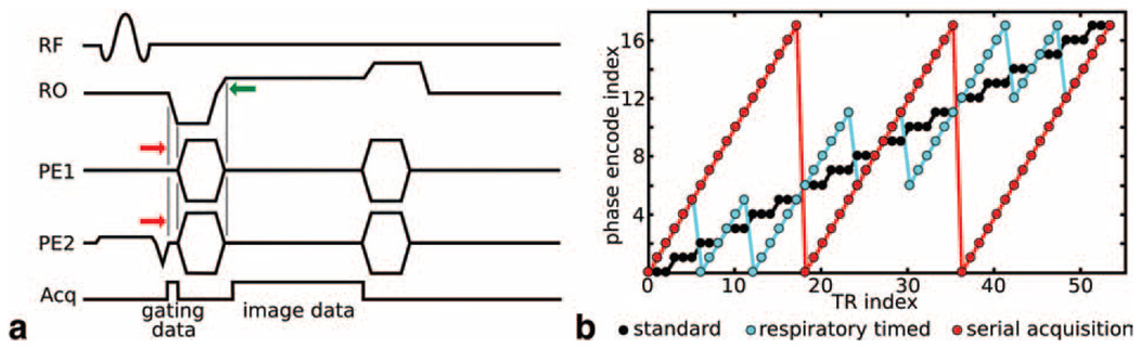
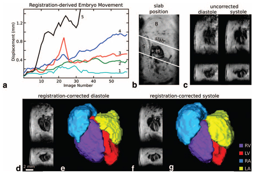
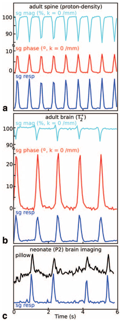
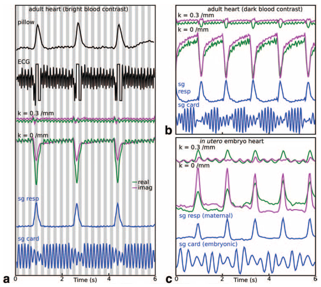
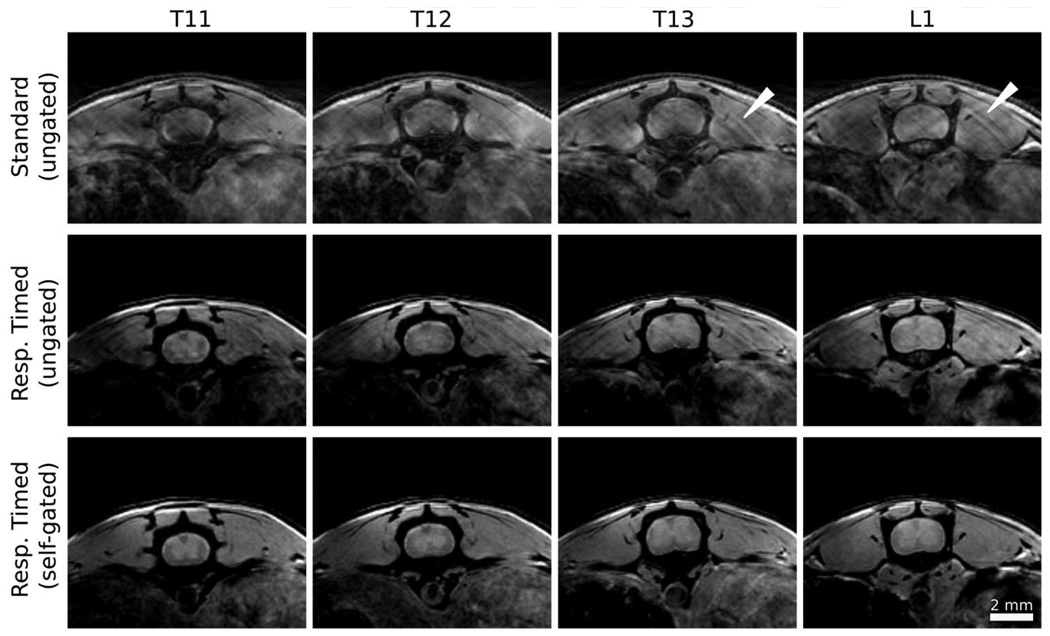
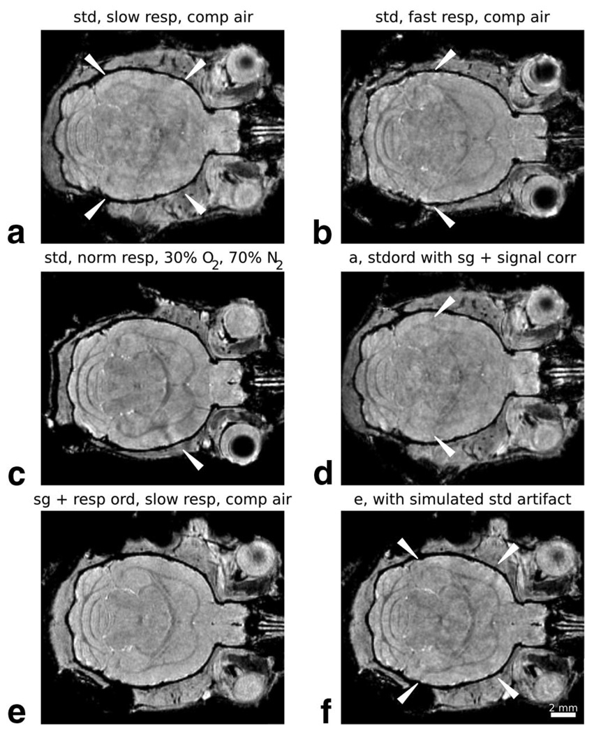
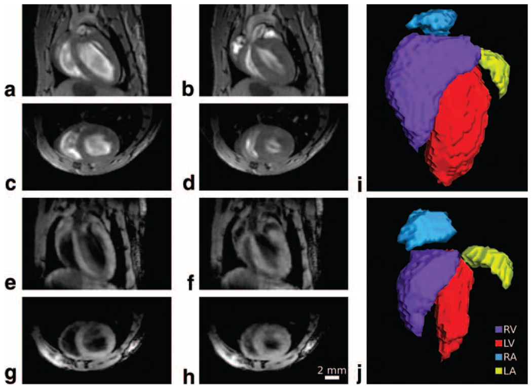
Motion during magnetic resonance imaging (MRI) scans routinely results in undesirable image artifact or blurring. Since high-resolution, three-dimensional (3D) imaging of the mouse requires long scan times for satisfactory signal-to-noise ratio (SNR) and image quality, motion-related artifacts are likely over much of the body and limit applications of mouse MRI. In this investigation, we explored the use of self-gated imaging methods and image coregistration for improving image quality in the presence of motion. Self-gated signal results from a modified 3D gradient-echo sequence showed detection of periodic respiratory and cardiac motion in the adult mouse-with excellent comparison to traditional measurements, sensitivity to respiration-induced tissue changes in the brain, and even detection of embryonic cardiac motion in utero. Serial image coregistration with rapidly-acquired, low-SNR volumes further enabled detection and correction of bulk changes in embryo location during in utero imaging sessions and subsequent reconstruction of high-quality images. These methods, in combination, are shown to expand the range of applications for 3D mouse MRI, enabling late-stage embryonic heart imaging and introducing the possibility of longitudinal developmental studies from embryonic stages through adulthood.
磁共振成像(MRI)扫描过程中的运动通常会导致不理想的图像伪影或模糊。由于对小鼠进行高分辨率三维(3D)成像需要较长的扫描时间以获得令人满意的信噪比(SNR)和图像质量,与运动相关的伪影很可能在身体的大部分区域出现,并限制了小鼠MRI的应用。在本研究中,我们探索了使用自门控成像方法和图像配准来在存在运动的情况下提高图像质量。来自改良3D梯度回波序列的自门控信号显示,可检测成年小鼠的周期性呼吸和心脏运动——与传统测量结果具有极佳的对比,对大脑中呼吸诱导的组织变化具有敏感性,甚至能检测子宫内胚胎的心脏运动。通过与快速采集的低SNR体积进行序列图像配准,进一步能够在子宫内成像过程中检测和校正胚胎位置的整体变化,并随后重建高质量图像。这些方法相结合,显示出可扩展3D小鼠MRI的应用范围,实现晚期胚胎心脏成像,并引入了从胚胎期到成年期进行纵向发育研究的可能性。