Beidler Stephanie K, Douillet Christelle D, Berndt Daniel F, Keagy Blair A, Rich Preston B, Marston William A
Department of Surgery, University of North Carolina School of Medicine, Chapel Hill, NC.
J Vasc Surg. 2009 Apr;49(4):1013-20. doi: 10.1016/j.jvs.2008.11.049.
Elevated inflammatory cytokine levels have been implicated in the pathogenesis of non-healing chronic venous insufficiency (CVI) ulcers. The goal of this study was to determine the protein levels of a wide range of inflammatory cytokines in untreated CVI ulcer tissue before and after 4 weeks of high-strength compression therapy. These levels were compared to cytokines present in healthy tissue.
Thirty limbs with untreated CVI and leg ulceration received therapy for 4 weeks with sustained high-compression bandaging at an ambulatory wound center. Biopsies were obtained from healthy and ulcerated tissue before and after therapy. A multiplexed protein assay was used to measure multiple cytokines in a single sample. Patients were designated as rapid or delayed healers based on ulcer surface area change.
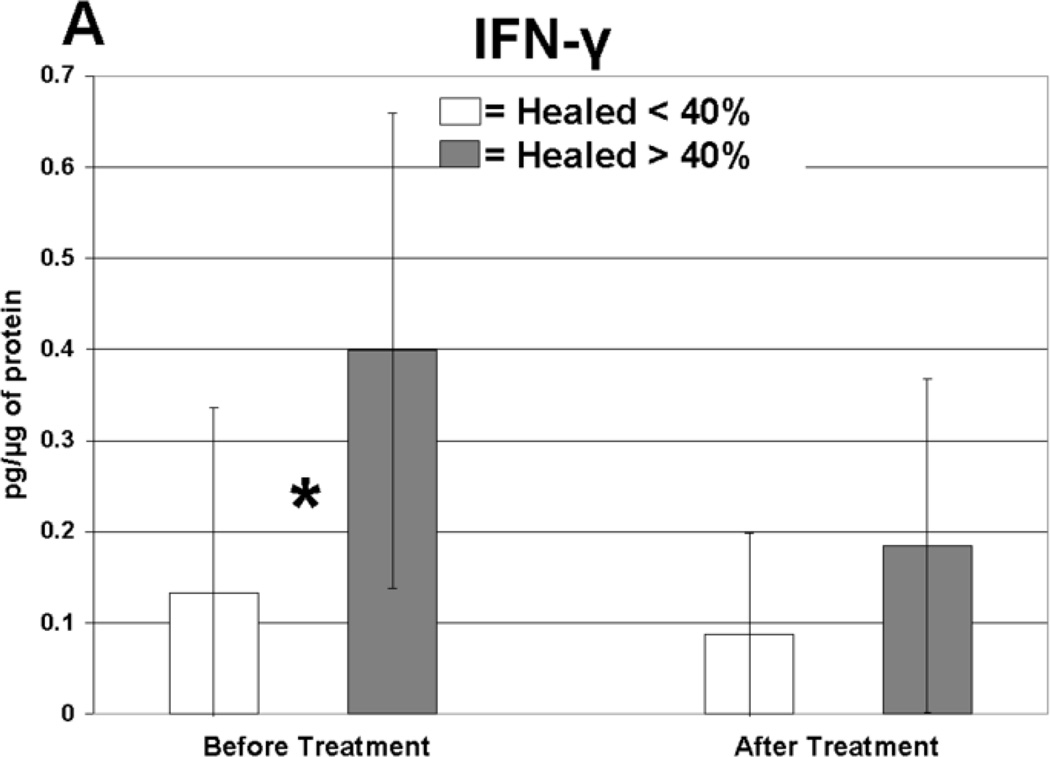
The majority of pro-inflammatory cytokine protein levels were elevated in ulcer tissue compared to healthy tissue, and compression therapy significantly reduced these cytokines. TGF-beta1 was upregulated in ulcer tissue following compression therapy. Rapid healing ulcers had significantly higher levels of IL-1alpha, IL-1beta, IFN-gamma, IL-12p40, and granulocyte macrophage colony stimulating factor (GM-CSF) before compression therapy, and IL-1 Ra after therapy. IFN-gamma levels significantly decreased following therapy in the rapidly healing patients.
CVI ulcer healing is associated with a pro-inflammatory environment prior to treatment that reflects metabolically active peri-wound tissue that has the potential to heal. Treatment with compression therapy results in healing that is coupled with reduced pro-inflammatory cytokine levels and higher levels of the anti-inflammatory cytokine IL-1 Ra.
炎症细胞因子水平升高与难愈合的慢性静脉功能不全(CVI)溃疡的发病机制有关。本研究的目的是确定高强度压迫治疗4周前后未经治疗的CVI溃疡组织中多种炎症细胞因子的蛋白水平。并将这些水平与健康组织中的细胞因子进行比较。
30例患有未经治疗的CVI和腿部溃疡的肢体在门诊伤口中心接受持续高压力绷带治疗4周。在治疗前后从健康组织和溃疡组织中获取活检样本。采用多重蛋白检测法在单个样本中测量多种细胞因子。根据溃疡表面积变化将患者分为愈合快或愈合慢的类型。
与健康组织相比,溃疡组织中大多数促炎细胞因子蛋白水平升高,压迫治疗可显著降低这些细胞因子水平。压迫治疗后溃疡组织中转化生长因子-β1上调。愈合快的溃疡在压迫治疗前白细胞介素-1α、白细胞介素-1β、干扰素-γ、白细胞介素-12p40和粒细胞巨噬细胞集落刺激因子(GM-CSF)水平显著较高,治疗后白细胞介素-1受体拮抗剂(IL-1 Ra)水平较高。在愈合快的患者中,治疗后干扰素-γ水平显著降低。
CVI溃疡愈合与治疗前的促炎环境有关,这反映了具有愈合潜力的代谢活跃的伤口周围组织。压迫治疗导致愈合的同时促炎细胞因子水平降低,抗炎细胞因子白细胞介素-1 Ra水平升高。