Shimada Kenichi, Chen Shuang, Dempsey Paul W, Sorrentino Rosalinda, Alsabeh Randa, Slepenkin Anatoly V, Peterson Ellena, Doherty Terence M, Underhill David, Crother Timothy R, Arditi Moshe
Division of Pediatrics, Infectious Diseases, and Immunology, Cedars-Sinai Medical Center, David Geffen School of Medicine, University of California Los Angeles, Los Angeles, CA, USA.
PLoS Pathog. 2009 Apr;5(4):e1000379. doi: 10.1371/journal.ppat.1000379. Epub 2009 Apr 10.
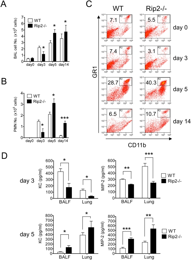
Here we investigated the role of the Nod/Rip2 pathway in host responses to Chlamydophila pneumoniae-induced pneumonia in mice. Rip2(-/-) mice infected with C. pneumoniae exhibited impaired iNOS expression and NO production, and delayed neutrophil recruitment to the lungs. Levels of IL-6 and IFN-gamma levels as well as KC and MIP-2 levels in bronchoalveolar lavage fluid (BALF) were significantly decreased in Rip2(-/-) mice compared to wild-type (WT) mice at day 3. Rip2(-/-) mice showed significant delay in bacterial clearance from the lungs and developed more severe and chronic lung inflammation that continued even on day 35 and led to increased mortality, whereas WT mice cleared the bacterial load, recovered from acute pneumonia, and survived. Both Nod1(-/-) and Nod2(-/-) mice also showed delayed bacterial clearance, suggesting that C. pneumoniae is recognized by both of these intracellular receptors. Bone marrow chimera experiments demonstrated that Rip2 in BM-derived cells rather than non-hematopoietic stromal cells played a key role in host responses in the lungs and clearance of C. pneumoniae. Furthermore, adoptive transfer of WT macrophages intratracheally was able to rescue the bacterial clearance defect in Rip2(-/-) mice. These results demonstrate that in addition to the TLR/MyD88 pathway, the Nod/Rip2 signaling pathway also plays a significant role in intracellular recognition, innate immune host responses, and ultimately has a decisive impact on clearance of C. pneumoniae from the lungs and survival of the infectious challenge.
在此,我们研究了Nod/Rip2信号通路在小鼠对肺炎衣原体诱导的肺炎的宿主反应中的作用。感染肺炎衣原体的Rip2基因敲除(-/-)小鼠表现出诱导型一氧化氮合酶(iNOS)表达和一氧化氮(NO)生成受损,以及中性粒细胞向肺部募集延迟。与野生型(WT)小鼠相比,在第3天时,Rip2(-/-)小鼠支气管肺泡灌洗液(BALF)中的白细胞介素-6(IL-6)和γ干扰素(IFN-γ)水平以及角质形成细胞趋化因子(KC)和巨噬细胞炎性蛋白-2(MIP-2)水平显著降低。Rip2(-/-)小鼠肺部细菌清除明显延迟,并出现更严重和慢性的肺部炎症,甚至在第35天时仍持续存在,导致死亡率增加,而WT小鼠清除了细菌负荷,从急性肺炎中恢复并存活下来。Nod1基因敲除(-/-)和Nod2基因敲除(-/-)小鼠也表现出细菌清除延迟,这表明肺炎衣原体可被这两种细胞内受体识别。骨髓嵌合体实验表明,骨髓来源细胞中的Rip2而非非造血基质细胞在肺部宿主反应和肺炎衣原体清除中起关键作用。此外,经气管内过继转移野生型巨噬细胞能够挽救Rip2(-/-)小鼠的细菌清除缺陷。这些结果表明,除了Toll样受体(TLR)/髓样分化因子88(MyD88)信号通路外,Nod/Rip2信号通路在细胞内识别、先天性免疫宿主反应中也发挥重要作用,并最终对肺部肺炎衣原体的清除和感染攻击后的存活产生决定性影响。